 ムービー
ムービー コントローラー


+ データを開く

- 基本情報
基本情報
| 登録情報 |  | |||||||||
|---|---|---|---|---|---|---|---|---|---|---|
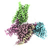
| タイトル | Cryo-EM structure of MrgD-Gi complex with beta-alanine | |||||||||
 マップデータ マップデータ | post-process map | |||||||||
 試料 試料 |
| |||||||||
| 機能・相同性 |  機能・相同性情報 機能・相同性情報Activation of the phototransduction cascade / Sensory perception of sweet, bitter, and umami (glutamate) taste / Olfactory Signaling Pathway / G beta:gamma signalling through PLC beta / Presynaptic function of Kainate receptors / Prostacyclin signalling through prostacyclin receptor / G alpha (z) signalling events / Glucagon-type ligand receptors / G beta:gamma signalling through PI3Kgamma / G beta:gamma signalling through CDC42 ...Activation of the phototransduction cascade / Sensory perception of sweet, bitter, and umami (glutamate) taste / Olfactory Signaling Pathway / G beta:gamma signalling through PLC beta / Presynaptic function of Kainate receptors / Prostacyclin signalling through prostacyclin receptor / G alpha (z) signalling events / Glucagon-type ligand receptors / G beta:gamma signalling through PI3Kgamma / G beta:gamma signalling through CDC42 / Synthesis, secretion, and inactivation of Glucagon-like Peptide-1 (GLP-1) / G beta:gamma signalling through BTK / Adrenaline,noradrenaline inhibits insulin secretion / ADP signalling through P2Y purinoceptor 12 / Cooperation of PDCL (PhLP1) and TRiC/CCT in G-protein beta folding / Thromboxane signalling through TP receptor / Thrombin signalling through proteinase activated receptors (PARs) / Activation of G protein gated Potassium channels / Inhibition of voltage gated Ca2+ channels via Gbeta/gamma subunits / G-protein activation / Ca2+ pathway / G alpha (s) signalling events / G alpha (q) signalling events / Extra-nuclear estrogen signaling / G alpha (12/13) signalling events / Vasopressin regulates renal water homeostasis via Aquaporins / G alpha (i) signalling events / GPER1 signaling / Glucagon-like Peptide-1 (GLP1) regulates insulin secretion / ADP signalling through P2Y purinoceptor 1 / phototransduction, visible light / alkylglycerophosphoethanolamine phosphodiesterase activity / photoreceptor outer segment membrane / spectrin binding / photoreceptor outer segment / Adenylate cyclase inhibitory pathway / positive regulation of protein localization to cell cortex / regulation of cAMP-mediated signaling / D2 dopamine receptor binding / G protein-coupled serotonin receptor binding / regulation of mitotic spindle organization / cellular response to forskolin / cardiac muscle cell apoptotic process / adenylate cyclase-inhibiting G protein-coupled receptor signaling pathway / photoreceptor inner segment / Regulation of insulin secretion / G protein-coupled receptor binding / G protein-coupled receptor activity / electron transport chain / G-protein beta/gamma-subunit complex binding / adenylate cyclase-modulating G protein-coupled receptor signaling pathway / ADP signalling through P2Y purinoceptor 12 / response to peptide hormone / Adrenaline,noradrenaline inhibits insulin secretion / G alpha (z) signalling events / ADORA2B mediated anti-inflammatory cytokines production / sensory perception of taste / GPER1 signaling / GDP binding / G-protein beta-subunit binding / heterotrimeric G-protein complex / signaling receptor complex adaptor activity / GTPase binding / myelin sheath / retina development in camera-type eye / phospholipase C-activating G protein-coupled receptor signaling pathway / cell cortex / midbody / positive regulation of cytosolic calcium ion concentration / cell body / G alpha (i) signalling events / fibroblast proliferation / cellular response to hypoxia / G alpha (s) signalling events / cell population proliferation / Extra-nuclear estrogen signaling / periplasmic space / electron transfer activity / iron ion binding / G protein-coupled receptor signaling pathway / cell cycle / cell division / lysosomal membrane / GTPase activity / centrosome / dendrite / heme binding / protein-containing complex binding / nucleolus / GTP binding / magnesium ion binding / extracellular space / extracellular exosome / nucleoplasm / plasma membrane / cytoplasm 類似検索 - 分子機能 | |||||||||
| 生物種 |  Homo sapiens (ヒト) / Homo sapiens (ヒト) /  | |||||||||
| 手法 | 単粒子再構成法 / クライオ電子顕微鏡法 / 解像度: 3.1 Å | |||||||||
 データ登録者 データ登録者 | Suzuki S / Iida M / Kawamoto A / Oshima A | |||||||||
| 資金援助 |  日本, 2件 日本, 2件
| |||||||||
 引用 引用 | ジャーナル: Commun Biol / 年: 2022 タイトル: Structural insight into the activation mechanism of MrgD with heterotrimeric Gi-protein revealed by cryo-EM. 著者: Shota Suzuki / Momoko Iida / Yoko Hiroaki / Kotaro Tanaka / Akihiro Kawamoto / Takayuki Kato / Atsunori Oshima / 要旨: MrgD, a member of the Mas-related G protein-coupled receptor (MRGPR) family, has high basal activity for Gi activation. It recognizes endogenous ligands, such as β-alanine, and is involved in pain ...MrgD, a member of the Mas-related G protein-coupled receptor (MRGPR) family, has high basal activity for Gi activation. It recognizes endogenous ligands, such as β-alanine, and is involved in pain and itch signaling. The lack of a high-resolution structure for MrgD hinders our understanding of whether its activation is ligand-dependent or constitutive. Here, we report two cryo-EM structures of the MrgD-Gi complex in the β-alanine-bound and apo states at 3.1 Å and 2.8 Å resolution, respectively. These structures show that β-alanine is bound to a shallow pocket at the extracellular domains. The extracellular half of the sixth transmembrane helix undergoes a significant movement and is tightly packed into the third transmembrane helix through hydrophobic residues, creating the active form. Our structures demonstrate a structural basis for the characteristic ligand recognition of MrgD. These findings provide a framework to guide drug designs targeting the MrgD receptor. | |||||||||
| 履歴 |
|
- 構造の表示
構造の表示
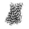
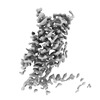
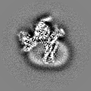
| 添付画像 |
|---|

- ダウンロードとリンク
-EMDBアーカイブ
| マップデータ | emd_33554.map.gz | 9.1 MB | EMDBマップデータ形式 | |
|---|---|---|---|---|
| ヘッダ (付随情報) | emd-33554-v30.xmlemd-33554.xml | 22.9 KB 22.9 KB | 表示 表示 | EMDBヘッダ |
| FSC (解像度算出) | emd_33554_fsc.xml | 11.4 KB | 表示 | FSCデータファイル |
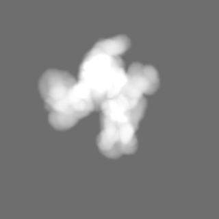
| 画像 | 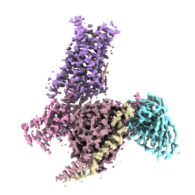 emd_33554.png emd_33554.png | 121 KB | ||
| マスクデータ | emd_33554_msk_1.map | 125 MB | マスクマップ | |
| その他 | emd_33554_half_map_1.map.gzemd_33554_half_map_2.map.gz | 98.4 MB 98.5 MB | ||
| アーカイブディレクトリ |  http://ftp.pdbj.org/pub/emdb/structures/EMD-33554ftp://ftp.pdbj.org/pub/emdb/structures/EMD-33554 http://ftp.pdbj.org/pub/emdb/structures/EMD-33554ftp://ftp.pdbj.org/pub/emdb/structures/EMD-33554 | HTTPS FTP |
-検証レポート
| 文書・要旨 | emd_33554_validation.pdf.gz | 712.6 KB | 表示 | EMDB検証レポート |
|---|---|---|---|---|
| 文書・詳細版 | emd_33554_full_validation.pdf.gz | 712.2 KB | 表示 | |
| XML形式データ | emd_33554_validation.xml.gz | 18.8 KB | 表示 | |
| CIF形式データ | emd_33554_validation.cif.gz | 24.6 KB | 表示 | |
| アーカイブディレクトリ | https://ftp.pdbj.org/pub/emdb/validation_reports/EMD-33554ftp://ftp.pdbj.org/pub/emdb/validation_reports/EMD-33554 | HTTPS FTP |
-関連構造データ
| 関連構造データ |  7y12MC  7y13C  7y14C  7y15C M: このマップから作成された原子モデル C: 同じ文献を引用 ( |
|---|---|
| 類似構造データ | |
| 電子顕微鏡画像生データ | EMPIAR-11073 (タイトル: Cryo-EM structure of MrgD-Gi complex with beta-alanine Data size: 4.1 TB Data #1: K3 movies for MrgD-Gi complex with beta-alanine [micrographs - multiframe]) |
-リンク
| EMDBのページ | EMDB (EBI/PDBe) / EMDataResource |
|---|---|
| 「今月の分子」の関連する項目 |


-マップ
| ファイル | ダウンロード / ファイル: emd_33554.map.gz / 形式: CCP4 / 大きさ: 125 MB / タイプ: IMAGE STORED AS FLOATING POINT NUMBER (4 BYTES) | ||||||||||||||||||||
|---|---|---|---|---|---|---|---|---|---|---|---|---|---|---|---|---|---|---|---|---|---|
| 注釈 | post-process map | ||||||||||||||||||||
| ボクセルのサイズ | X=Y=Z: 0.675 Å | ||||||||||||||||||||
| 密度 |
| ||||||||||||||||||||
| 対称性 | 空間群: 1 | ||||||||||||||||||||
| 詳細 | EMDB XML:
|
-添付データ
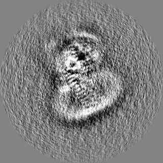
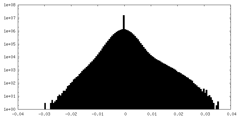
-マスク #1
| ファイル | emd_33554_msk_1.map | ||||||||||||
|---|---|---|---|---|---|---|---|---|---|---|---|---|---|
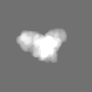
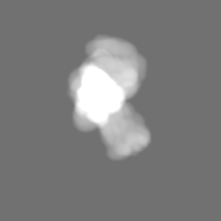
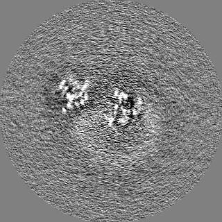
| 投影像・断面図 |
| ||||||||||||
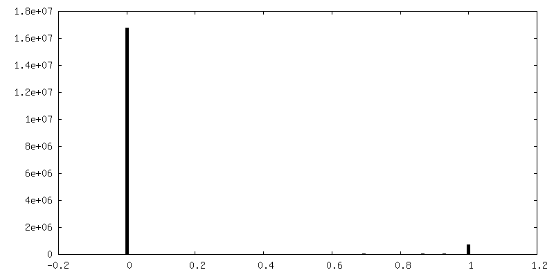
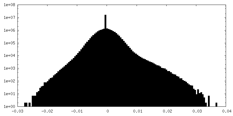
| 密度ヒストグラム |
 Z
Z Y
Y X
X


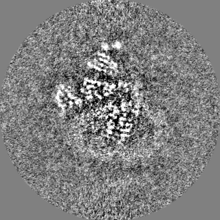
-ハーフマップ: half map 1
| ファイル | emd_33554_half_map_1.map | ||||||||||||
|---|---|---|---|---|---|---|---|---|---|---|---|---|---|
| 注釈 | half map 1 | ||||||||||||
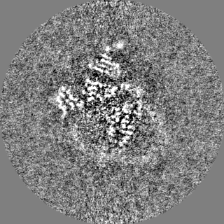
| 投影像・断面図 |
| ||||||||||||
| 密度ヒストグラム |


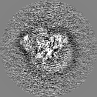
-ハーフマップ: half map 2
| ファイル | emd_33554_half_map_2.map | ||||||||||||
|---|---|---|---|---|---|---|---|---|---|---|---|---|---|
| 注釈 | half map 2 | ||||||||||||
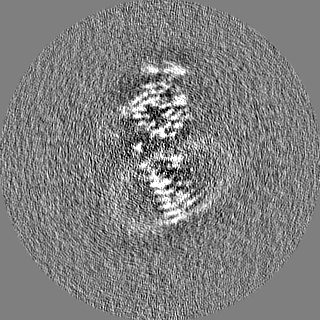
| 投影像・断面図 |
| ||||||||||||
| 密度ヒストグラム |


- 試料の構成要素
+全体 : beta-alanine bound MrgD-Gi complex with the scFv16
+超分子 #1: beta-alanine bound MrgD-Gi complex with the scFv16
+超分子 #2: G(i) subunit alpha-1,Mas-related G-protein coupled receptor member D
+超分子 #3: subunit beta-1,subunit gamma-2
+超分子 #4: scFV16
+分子 #1: Guanine nucleotide-binding protein G(i) subunit alpha-1

 Spodoptera frugiperda (ツマジロクサヨトウ)
+分子 #2: Guanine nucleotide-binding protein G(I)/G(S)/G(T) subunit beta-1
+分子 #3: Guanine nucleotide-binding protein G(I)/G(S)/G(O) subunit gamma-2
+分子 #4: Soluble cytochrome b562,Mas-related G-protein coupled receptor me...
+分子 #5: scFV16
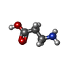
+分子 #6: BETA-ALANINE
+分子 #7: PALMITIC ACID

-実験情報
-構造解析
| 手法 | クライオ電子顕微鏡法 |
|---|---|
 解析 解析 | 単粒子再構成法 |
| 試料の集合状態 | particle |
-試料調製
| 濃度 | 10 mg/mL |
|---|---|
| 緩衝液 | pH: 7.4 |
| グリッド | モデル: Quantifoil R1.2/1.3 / 材質: GOLD / メッシュ: 300 / 支持フィルム - 材質: CARBON / 支持フィルム - トポロジー: HOLEY ARRAY / 前処理 - タイプ: GLOW DISCHARGE / 前処理 - 時間: 60 sec. |
| 凍結 | 凍結剤: ETHANE / チャンバー内湿度: 100 % / チャンバー内温度: 277 K / 装置: FEI VITROBOT MARK IV / 詳細: blot time 3 seconds blot force 5. |
- 電子顕微鏡法
| 顕微鏡 | FEI TITAN KRIOS |
|---|---|
| 特殊光学系 | 球面収差補正装置: The Microscope implicated Cs corrector. |
| 撮影 | フィルム・検出器のモデル: GATAN K3 BIOQUANTUM (6k x 4k) 平均電子線量: 60.0 e/Å2 |
| 電子線 | 加速電圧: 300 kV / 電子線源:  FIELD EMISSION GUN FIELD EMISSION GUN |
| 電子光学系 | 照射モード: FLOOD BEAM / 撮影モード: BRIGHT FIELD / Cs: 0.01 mm 最大 デフォーカス(公称値): 1.9000000000000001 µm 最小 デフォーカス(公称値): 0.7000000000000001 µm |
| 試料ステージ | 試料ホルダーモデル: FEI TITAN KRIOS AUTOGRID HOLDER ホルダー冷却材: NITROGEN |
| 実験機器 |  モデル: Titan Krios / 画像提供: FEI Company |
-画像解析
| 最終 再構成 | 想定した対称性 - 点群: C1 (非対称) / 解像度のタイプ: BY AUTHOR / 解像度: 3.1 Å / 解像度の算出法: FSC 0.143 CUT-OFF / 使用した粒子像数: 97282 |
|---|---|
| 初期 角度割当 | タイプ: MAXIMUM LIKELIHOOD |
| 最終 角度割当 | タイプ: MAXIMUM LIKELIHOOD |
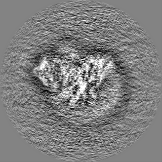
| FSC曲線 (解像度の算出) |  |
