 Movie
Movie Controller

[English] 日本語
 Yorodumi
- EMDB-33496: Tomographic reconstruction of FIB-milled cryo-lamella of HepG2 ce... -
+ Open data

- Basic information
| Entry |  | |||||||||
|---|---|---|---|---|---|---|---|---|---|---|
| Title | Tomographic reconstruction of FIB-milled cryo-lamella of HepG2 cells containing a LD-mitochondria contact sites. | |||||||||
 Map data Map data | ||||||||||
 Sample Sample |
| |||||||||
| Biological species |  Homo sapiens (human) Homo sapiens (human) | |||||||||
| Method | electron tomography / cryo EM | |||||||||
 Authors Authors | Xiao K / Li W / Li Z / Guo Q / Tao X / Ji W | |||||||||
| Funding support |  China, 1 items China, 1 items
| |||||||||
 Citation Citation | Journal: To Be Published Title: The functional universe of membrane contact sites. Authors: Prinz WA / Toulmay A / Balla T | |||||||||
| History |
|
- Structure visualization
| Supplemental images |
|---|

- Downloads & links
-EMDB archive
| Map data | emd_33496.map.gz | 628.8 MB |  EMDB map data format EMDB map data format | |
|---|---|---|---|---|
| Header (meta data) | emd-33496-v30.xmlemd-33496.xml | 7.3 KB 7.3 KB | Display Display | EMDB header |
| Images | 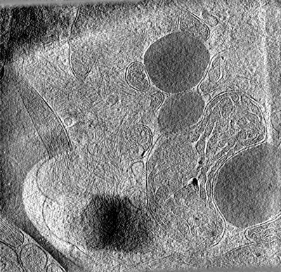 emd_33496.png emd_33496.png | 261.7 KB | ||
| Archive directory |  http://ftp.pdbj.org/pub/emdb/structures/EMD-33496ftp://ftp.pdbj.org/pub/emdb/structures/EMD-33496 http://ftp.pdbj.org/pub/emdb/structures/EMD-33496ftp://ftp.pdbj.org/pub/emdb/structures/EMD-33496 | HTTPS FTP |
-Validation report
| Summary document | emd_33496_validation.pdf.gz | 400.5 KB | Display | EMDB validaton report |
|---|---|---|---|---|
| Full document | emd_33496_full_validation.pdf.gz | 400 KB | Display | |
| Data in XML | emd_33496_validation.xml.gz | 5.3 KB | Display | |
| Data in CIF | emd_33496_validation.cif.gz | 5.8 KB | Display | |
| Arichive directory | https://ftp.pdbj.org/pub/emdb/validation_reports/EMD-33496ftp://ftp.pdbj.org/pub/emdb/validation_reports/EMD-33496 | HTTPS FTP |
-Links
| EMDB pages | EMDB (EBI/PDBe) / EMDataResource |
|---|
-Map
| File | Download / File: emd_33496.map.gz / Format: CCP4 / Size: 679.7 MB / Type: IMAGE STORED AS FLOATING POINT NUMBER (4 BYTES) | ||||||||||||||||||||
|---|---|---|---|---|---|---|---|---|---|---|---|---|---|---|---|---|---|---|---|---|---|
| Voxel size | X=Y=Z: 21.68 Å | ||||||||||||||||||||
| Density |
| ||||||||||||||||||||
| Symmetry | Space group: 1 | ||||||||||||||||||||
| Details | EMDB XML:
|
-Supplemental data
- Sample components
-Entire : HepG2 cells with fluorescent reporter of LD-mitochondria contact
| Entire | Name: HepG2 cells with fluorescent reporter of LD-mitochondria contact |
|---|---|
| Components |
|
-Supramolecule #1: HepG2 cells with fluorescent reporter of LD-mitochondria contact
| Supramolecule | Name: HepG2 cells with fluorescent reporter of LD-mitochondria contact type: cell / ID: 1 / Parent: 0 |
|---|---|
| Source (natural) | Organism: Homo sapiens (human) |
-Experimental details
-Structure determination
| Method | cryo EM |
|---|---|
 Processing Processing | electron tomography |
| Aggregation state | cell |
-Sample preparation
| Buffer | pH: 7.4 |
|---|---|
| Vitrification | Cryogen name: ETHANE / Chamber humidity: 85 % / Chamber temperature: 310 K |
| Sectioning | Focused ion beam - Instrument: OTHER / Focused ion beam - Ion: OTHER / Focused ion beam - Voltage: 30 kV / Focused ion beam - Current: 0.5 nA / Focused ion beam - Duration: 300 sec. / Focused ion beam - Temperature: 303 K / Focused ion beam - Initial thickness: 1000 nm / Focused ion beam - Final thickness: 150 nm Focused ion beam - Details: The value given for _em_focused_ion_beam.instrument is Tescan S8000G. This is not in a list of allowed values {'DB235', 'OTHER'} so OTHER is written into the XML file. |
- Electron microscopy
| Microscope | FEI TITAN KRIOS |
|---|---|
| Image recording | Film or detector model: GATAN K2 QUANTUM (4k x 4k) / Average electron dose: 3.0 e/Å2 |
| Electron beam | Acceleration voltage: 300 kV / Electron source:  FIELD EMISSION GUN FIELD EMISSION GUN |
| Electron optics | Illumination mode: FLOOD BEAM / Imaging mode: BRIGHT FIELD / Nominal defocus max: 6.0 µm / Nominal defocus min: 5.0 µm |
| Experimental equipment |  Model: Titan Krios / Image courtesy: FEI Company |
-Image processing
| Final reconstruction | Algorithm: BACK PROJECTION / Number images used: 35 |
|---|
