 Movie
Movie Controller


+ Open data

- Basic information
| Entry | Database: EMDB / ID: EMD-3345 | |||||||||
|---|---|---|---|---|---|---|---|---|---|---|
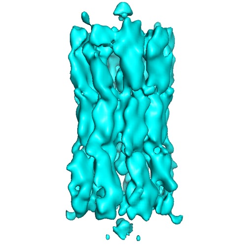
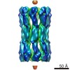
| Title | Hexadecameric structure of an invertebrate gap junction channel | |||||||||
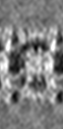
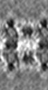
 Map data Map data | This is a map showing a single INX-6 gap junction channel with P2 crystallographic symmetry | |||||||||
 Sample Sample |
| |||||||||
 Keywords Keywords | innexin / gap junction channel / cryo-electron crystallography / three-dimensional reconstruction / two-dimensional crystal | |||||||||
| Biological species |  | |||||||||
| Method | electron crystallography / cryo EM / Resolution: 10.0 Å | |||||||||
 Authors Authors | Oshima A / Matsuzawa T / Murata K / Tani K / Fujiyoshi Y | |||||||||
 Citation Citation | Journal: J Mol Biol / Year: 2016 Title: Hexadecameric structure of an invertebrate gap junction channel. Authors: Atsunori Oshima / Tomohiro Matsuzawa / Kazuyoshi Murata / Kazutoshi Tani / Yoshinori Fujiyoshi /  Abstract: Innexins are invertebrate-specific gap junction proteins with four transmembrane helices. These proteins oligomerize to constitute intercellular channels that allow for the passage of small signaling ...Innexins are invertebrate-specific gap junction proteins with four transmembrane helices. These proteins oligomerize to constitute intercellular channels that allow for the passage of small signaling molecules associated with neural and muscular electrical activity. In contrast to the large number of structural and functional studies of connexin gap junction channels, few structural studies of recombinant innexin channels are reported. Here we show the three-dimensional structure of two-dimensionally crystallized Caenorhabditis elegans innexin-6 (INX-6) gap junction channels. The N-terminal deleted INX-6 proteins are crystallized in lipid bilayers. The three-dimensional reconstruction determined by cryo-electron crystallography reveals that a single INX-6 gap junction channel comprises 16 subunits, a hexadecamer, in contrast to chordate connexin channels, which comprise 12 subunits. The channel pore diameters at the cytoplasmic entrance and extracellular gap region are larger than those of connexin26. Two bulb densities are observed in each hemichannel, one in the pore and the other at the cytoplasmic side of the hemichannel in the channel pore pathway. These findings imply a structural diversity of gap junction channels among multicellular organisms. | |||||||||
| History |
|
- Structure visualization
| Movie |
 Movie viewer Movie viewer |
|---|---|
| Structure viewer | EM map: SurfViewMolmilJmol/JSmol |
| Supplemental images |
- Downloads & links
-EMDB archive
| Map data | emd_3345.map.gz | 809 KB | EMDB map data format | |
|---|---|---|---|---|
| Header (meta data) | emd-3345-v30.xmlemd-3345.xml | 10.8 KB 10.8 KB | Display Display | EMDB header |
| Images | EMD-3345.tif | 168.1 KB | ||
| Archive directory |  http://ftp.pdbj.org/pub/emdb/structures/EMD-3345ftp://ftp.pdbj.org/pub/emdb/structures/EMD-3345 http://ftp.pdbj.org/pub/emdb/structures/EMD-3345ftp://ftp.pdbj.org/pub/emdb/structures/EMD-3345 | HTTPS FTP |
-Validation report
| Summary document | emd_3345_validation.pdf.gz | 199.8 KB | Display | EMDB validaton report |
|---|---|---|---|---|
| Full document | emd_3345_full_validation.pdf.gz | 198.9 KB | Display | |
| Data in XML | emd_3345_validation.xml.gz | 4.5 KB | Display | |
| Arichive directory | https://ftp.pdbj.org/pub/emdb/validation_reports/EMD-3345ftp://ftp.pdbj.org/pub/emdb/validation_reports/EMD-3345 | HTTPS FTP |
-Related structure data
-Links
| EMDB pages | EMDB (EBI/PDBe) / EMDataResource |
|---|
-Map
| File | Download / File: emd_3345.map.gz / Format: CCP4 / Size: 2.1 MB / Type: IMAGE STORED AS FLOATING POINT NUMBER (4 BYTES) | ||||||||||||||||||||||||||||||||||||||||||||||||||||||||||||||||||||
|---|---|---|---|---|---|---|---|---|---|---|---|---|---|---|---|---|---|---|---|---|---|---|---|---|---|---|---|---|---|---|---|---|---|---|---|---|---|---|---|---|---|---|---|---|---|---|---|---|---|---|---|---|---|---|---|---|---|---|---|---|---|---|---|---|---|---|---|---|---|
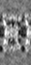
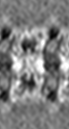
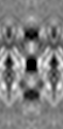
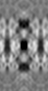
| Annotation | This is a map showing a single INX-6 gap junction channel with P2 crystallographic symmetry | ||||||||||||||||||||||||||||||||||||||||||||||||||||||||||||||||||||
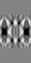
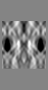
| Projections & slices | Image control
Images are generated by Spider. generated in cubic-lattice coordinate | ||||||||||||||||||||||||||||||||||||||||||||||||||||||||||||||||||||
| Voxel size | X: 2.4397 Å / Y: 2.5177 Å / Z: 2.5 Å | ||||||||||||||||||||||||||||||||||||||||||||||||||||||||||||||||||||
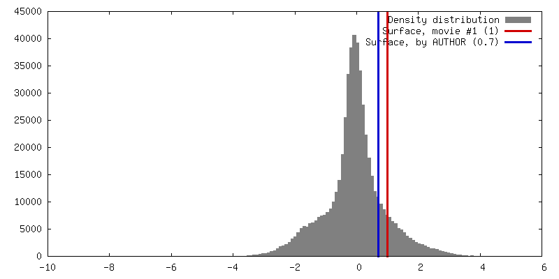
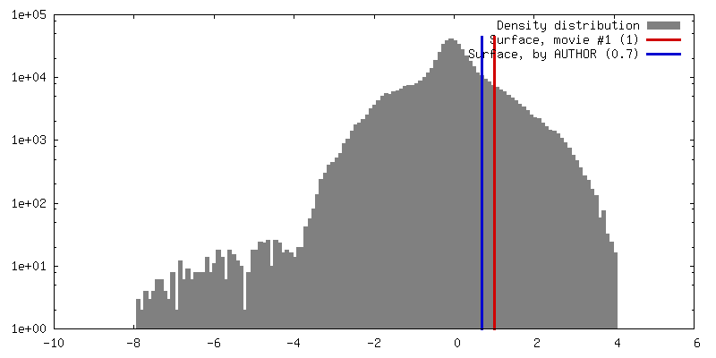
| Density |
| ||||||||||||||||||||||||||||||||||||||||||||||||||||||||||||||||||||
| Symmetry | Space group: 1 | ||||||||||||||||||||||||||||||||||||||||||||||||||||||||||||||||||||
| Details | EMDB XML:
CCP4 map header:
| ||||||||||||||||||||||||||||||||||||||||||||||||||||||||||||||||||||
 Z (Sec.)
 X (Row.)
 Y (Col.)


-Supplemental data
- Sample components
-Entire : The N-terminal deleted C. elegans innexin-6
| Entire | Name: The N-terminal deleted C. elegans innexin-6 |
|---|---|
| Components |
|
-Supramolecule #1000: The N-terminal deleted C. elegans innexin-6
| Supramolecule | Name: The N-terminal deleted C. elegans innexin-6 / type: sample / ID: 1000 / Details: The sample was reconstituted in lipid bilayers. / Oligomeric state: hexadecamer / Number unique components: 1 |
|---|---|
| Molecular weight | Experimental: 700 KDa / Theoretical: 700 KDa / Method: MALDI-TOF |
-Macromolecule #1: innexin-6
| Macromolecule | Name: innexin-6 / type: protein_or_peptide / ID: 1 / Name.synonym: INX-6 / Number of copies: 16 / Oligomeric state: 16 / Recombinant expression: Yes |
|---|---|
| Source (natural) | Organism: |
| Molecular weight | Experimental: 700 KDa / Theoretical: 700 KDa |
| Recombinant expression | Organism:   Spodoptera frugiperda (fall armyworm) / Recombinant cell: Sf9 / Recombinant plasmid: pFastbac Spodoptera frugiperda (fall armyworm) / Recombinant cell: Sf9 / Recombinant plasmid: pFastbac |
-Experimental details
-Structure determination
| Method | cryo EM |
|---|---|
 Processing Processing | electron crystallography |
| Aggregation state | 2D array |
-Sample preparation
| Concentration | 0.5 mg/mL |
|---|---|
| Buffer | pH: 7.5 / Details: 10 mM Tris (pH 7.5), 500 mM NaCl, and 1 mM EDTA |
| Vitrification | Cryogen name: ETHANE / Chamber temperature: 120 K / Instrument: FEI VITROBOT MARK IV / Method: Blot for 30 seconds before plunging |
| Details | Dialysis |
| Crystal formation | Details: Dialysis |
- Electron microscopy
| Microscope | JEOL KYOTO-3000SFF |
|---|---|
| Temperature | Min: 4 K / Max: 20 K / Average: 4 K |
| Date | Jul 26, 2014 |
| Image recording | Category: FILM / Film or detector model: KODAK SO-163 FILM / Digitization - Scanner: ZEISS SCAI / Digitization - Sampling interval: 7 µm / Number real images: 249 / Average electron dose: 20 e/Å2 / Camera length: 2000 / Bits/pixel: 8 |
| Tilt angle min | 0 |
| Electron beam | Acceleration voltage: 300 kV / Electron source:  FIELD EMISSION GUN FIELD EMISSION GUN |
| Electron optics | Calibrated magnification: 38210 / Illumination mode: FLOOD BEAM / Imaging mode: BRIGHT FIELD / Cs: 1.6 mm / Nominal defocus max: 3.459 µm / Nominal defocus min: 0.66 µm / Nominal magnification: 40000 |
| Sample stage | Specimen holder model: JEOL / Tilt angle max: 45 / Tilt series - Axis1 - Min angle: 0 ° / Tilt series - Axis1 - Max angle: 45 ° |
-Image processing
| Details | Images were processed with the MRC 2D crystal processing package. |
|---|---|
| Final reconstruction | Resolution.type: BY AUTHOR / Resolution: 10.0 Å / Resolution method: DIFFRACTION PATTERN/LAYERLINES / Software - Name: MRC |
| Crystal parameters | Unit cell - A: 118.5 Å / Unit cell - B: 111.5 Å / Unit cell - C: 320 Å / Unit cell - γ: 121.7 ° / Unit cell - α: 90.0 ° / Unit cell - β: 90.0 ° / Plane group: P 2 |
| CTF correction | Details: Each micrograph |
