 Movie
Movie Controller

[English] 日本語
 Yorodumi
- EMDB-33417: Microtubule triplets with complete C tubule of human centrioles f... -
+ Open data

- Basic information
| Entry |  | |||||||||
|---|---|---|---|---|---|---|---|---|---|---|
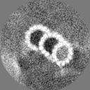
| Title | Microtubule triplets with complete C tubule of human centrioles from HeLa cells. | |||||||||
 Map data Map data | Postprocessed map of microtubule triplets with complete C tubule of human centrioles from HeLa cells. | |||||||||
 Sample Sample |
| |||||||||
 Keywords Keywords | Complex / Centriole / Microtubule / in situ / CELL CYCLE | |||||||||
| Biological species |  Homo sapiens (human) Homo sapiens (human) | |||||||||
| Method | subtomogram averaging / cryo EM / Resolution: 31.0 Å | |||||||||
 Authors Authors | Li SG / Wang ZY / Zhu Y / Ji G / Sun F | |||||||||
| Funding support |  China, 1 items China, 1 items
| |||||||||
 Citation Citation | Journal: Nat.Methods / Year: 2023 Title: ELI trifocal microscope: a precise system to prepare target cryo-lamellae for in situ cryo-ET study Authors: Li SG / Wang ZY / Jia X / Niu TX / Zhang JG / Yin GL / Zhang XY / Zhu Y / Ji G / Sun F | |||||||||
| History |
|
- Structure visualization
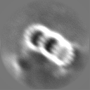
| Supplemental images |
|---|

- Downloads & links
-EMDB archive
| Map data | emd_33417.map.gz | 985.6 KB |  EMDB map data format EMDB map data format | |
|---|---|---|---|---|
| Header (meta data) | emd-33417-v30.xmlemd-33417.xml | 12.4 KB 12.4 KB | Display Display | EMDB header |
| FSC (resolution estimation) | emd_33417_fsc.xml | 4.7 KB | Display | FSC data file |
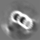
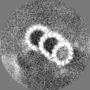
| Images | 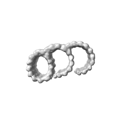 emd_33417.png emd_33417.png | 24.4 KB | ||
| Masks | emd_33417_msk_1.map | 8 MB | Mask map | |
| Filedesc metadata | emd-33417.cif.gz | 3.8 KB | ||
| Others | emd_33417_half_map_1.map.gzemd_33417_half_map_2.map.gz | 6 MB 6 MB | ||
| Archive directory |  http://ftp.pdbj.org/pub/emdb/structures/EMD-33417ftp://ftp.pdbj.org/pub/emdb/structures/EMD-33417 http://ftp.pdbj.org/pub/emdb/structures/EMD-33417ftp://ftp.pdbj.org/pub/emdb/structures/EMD-33417 | HTTPS FTP |
-Related structure data


-Links
| EMDB pages | EMDB (EBI/PDBe) / EMDataResource |
|---|
-Map
| File | Download / File: emd_33417.map.gz / Format: CCP4 / Size: 8 MB / Type: IMAGE STORED AS FLOATING POINT NUMBER (4 BYTES) | ||||||||||||||||||||||||||||||||||||
|---|---|---|---|---|---|---|---|---|---|---|---|---|---|---|---|---|---|---|---|---|---|---|---|---|---|---|---|---|---|---|---|---|---|---|---|---|---|
| Annotation | Postprocessed map of microtubule triplets with complete C tubule of human centrioles from HeLa cells. | ||||||||||||||||||||||||||||||||||||
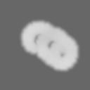
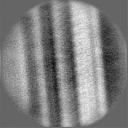
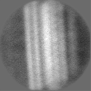
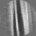
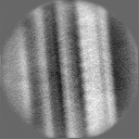
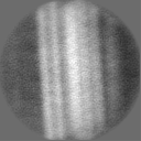
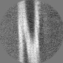
| Projections & slices | Image control
Images are generated by Spider. | ||||||||||||||||||||||||||||||||||||
| Voxel size | X=Y=Z: 8.6 Å | ||||||||||||||||||||||||||||||||||||
| Density |
| ||||||||||||||||||||||||||||||||||||
| Symmetry | Space group: 1 | ||||||||||||||||||||||||||||||||||||
| Details | EMDB XML:
|
 Z (Sec.)
 Y (Row.)
 X (Col.)


-Supplemental data
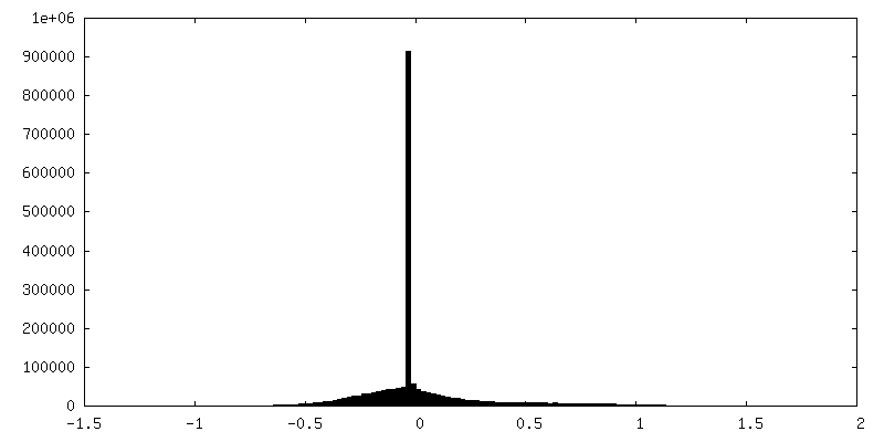
-Mask #1
| File | emd_33417_msk_1.map | ||||||||||||
|---|---|---|---|---|---|---|---|---|---|---|---|---|---|
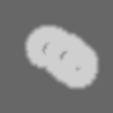
| Projections & Slices |
| ||||||||||||
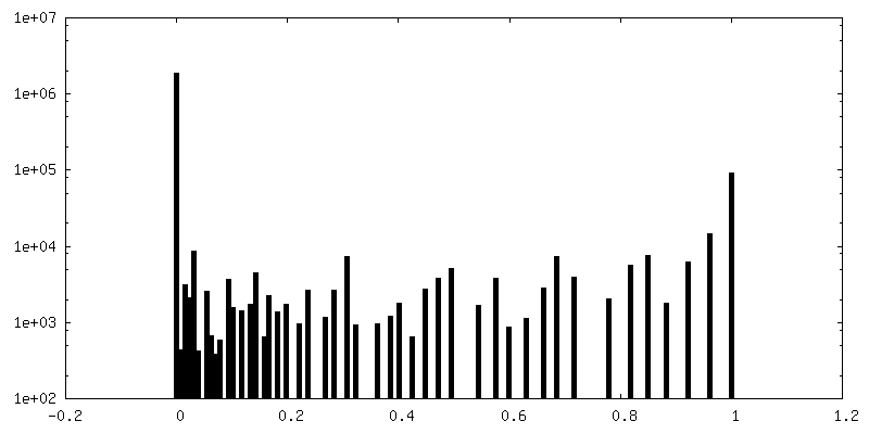
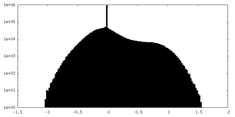
| Density Histograms |


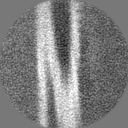
-Half map: Half map of microtubule triplets with complete C...
| File | emd_33417_half_map_1.map | ||||||||||||
|---|---|---|---|---|---|---|---|---|---|---|---|---|---|
| Annotation | Half map of microtubule triplets with complete C tubule of human centrioles from HeLa cells. | ||||||||||||
| Projections & Slices |
| ||||||||||||
| Density Histograms |

-Half map: Half map of microtubule triplets with complete C...
| File | emd_33417_half_map_2.map | ||||||||||||
|---|---|---|---|---|---|---|---|---|---|---|---|---|---|
| Annotation | Half map of microtubule triplets with complete C tubule of human centrioles from HeLa cells. | ||||||||||||
| Projections & Slices |
| ||||||||||||
| Density Histograms |


- Sample components
-Entire : Microtubule triplets with complete C tubule of human centrioles f...
| Entire | Name: Microtubule triplets with complete C tubule of human centrioles from HeLa cells. |
|---|---|
| Components |
|
-Supramolecule #1: Microtubule triplets with complete C tubule of human centrioles f...
| Supramolecule | Name: Microtubule triplets with complete C tubule of human centrioles from HeLa cells. type: organelle_or_cellular_component / ID: 1 / Parent: 0 |
|---|---|
| Source (natural) | Organism: Homo sapiens (human) |
-Experimental details
-Structure determination
| Method | cryo EM |
|---|---|
 Processing Processing | subtomogram averaging |
| Aggregation state | cell |
-Sample preparation
| Buffer | pH: 7 |
|---|---|
| Vitrification | Cryogen name: ETHANE |
- Electron microscopy
| Microscope | FEI TITAN KRIOS |
|---|---|
| Image recording | Film or detector model: GATAN K2 QUANTUM (4k x 4k) / Average electron dose: 3.0 e/Å2 |
| Electron beam | Acceleration voltage: 300 kV / Electron source:  FIELD EMISSION GUN FIELD EMISSION GUN |
| Electron optics | Illumination mode: SPOT SCAN / Imaging mode: BRIGHT FIELD / Cs: 2.7 mm / Nominal defocus max: 0.9 µm / Nominal defocus min: 0.5 µm |
| Experimental equipment |  Model: Titan Krios / Image courtesy: FEI Company |
-Image processing
| Final reconstruction | Applied symmetry - Point group: C1 (asymmetric) / Resolution.type: BY AUTHOR / Resolution: 31.0 Å / Resolution method: FSC 0.143 CUT-OFF / Number subtomograms used: 1762 |
|---|---|
| Extraction | Number tomograms: 59 / Number images used: 15939 |
| Final angle assignment | Type: MAXIMUM LIKELIHOOD |
| FSC plot (resolution estimation) | 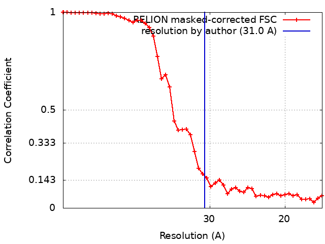 |
