 Movie
Movie Controller

[English] 日本語
 Yorodumi
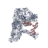
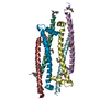
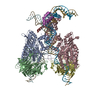
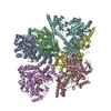
- EMDB-3333: Structure of a Group II Intron Complexed with its Reverse Transcr... -
+ Open data

- Basic information
| Entry | Database: EMDB / ID: EMD-3333 | |||||||||
|---|---|---|---|---|---|---|---|---|---|---|
| Title | Structure of a Group II Intron Complexed with its Reverse Transcriptase | |||||||||
 Map data Map data | Structure a of Group II Intron Complexed with its Reverse Transcriptase, Without Mask | |||||||||
 Sample Sample |
| |||||||||
| Function / homology |  Function and homology information Function and homology informationintron homing / RNA-directed DNA polymerase / mRNA processing / RNA-directed DNA polymerase activity / endonuclease activity / Hydrolases; Acting on ester bonds Similarity search - Function | |||||||||
| Biological species |  Lactococcus lactis (lactic acid bacteria) Lactococcus lactis (lactic acid bacteria) | |||||||||
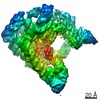
| Method | single particle reconstruction / cryo EM / Resolution: 4.2 Å | |||||||||
 Authors Authors | Qu G / Kaushal PS / Wang J / Shigematsu H / Piazza CL / Agrawal RK / Belfort M / Wang HW | |||||||||
 Citation Citation | Journal: Nat Struct Mol Biol / Year: 2016 Title: Structure of a group II intron in complex with its reverse transcriptase. Authors: Guosheng Qu / Prem Singh Kaushal / Jia Wang / Hideki Shigematsu / Carol Lyn Piazza / Rajendra Kumar Agrawal / Marlene Belfort / Hong-Wei Wang /   Abstract: Bacterial group II introns are large catalytic RNAs related to nuclear spliceosomal introns and eukaryotic retrotransposons. They self-splice, yielding mature RNA, and integrate into DNA as ...Bacterial group II introns are large catalytic RNAs related to nuclear spliceosomal introns and eukaryotic retrotransposons. They self-splice, yielding mature RNA, and integrate into DNA as retroelements. A fully active group II intron forms a ribonucleoprotein complex comprising the intron ribozyme and an intron-encoded protein that performs multiple activities including reverse transcription, in which intron RNA is copied into the DNA target. Here we report cryo-EM structures of an endogenously spliced Lactococcus lactis group IIA intron in its ribonucleoprotein complex form at 3.8-Å resolution and in its protein-depleted form at 4.5-Å resolution, revealing functional coordination of the intron RNA with the protein. Remarkably, the protein structure reveals a close relationship between the reverse transcriptase catalytic domain and telomerase, whereas the active splicing center resembles the spliceosomal Prp8 protein. These extraordinary similarities hint at intricate ancestral relationships and provide new insights into splicing and retromobility. | |||||||||
| History |
|
- Structure visualization
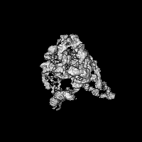
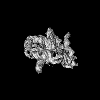
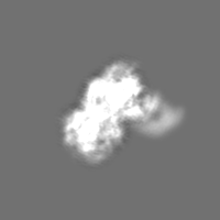
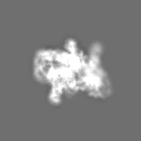
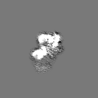
| Movie |
Movie viewer |
|---|---|
| Structure viewer | EM map: SurfViewMolmilJmol/JSmol |
| Supplemental images |

- Downloads & links
-EMDB archive
| Map data | emd_3333.map.gz | 3.6 MB | EMDB map data format | |
|---|---|---|---|---|
| Header (meta data) | emd-3333-v30.xmlemd-3333.xml | 7.9 KB 7.9 KB | Display Display | EMDB header |
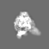
| Images | 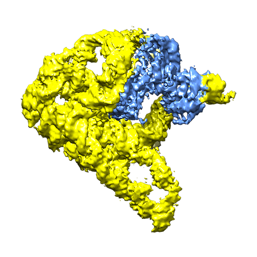 EMD-3333.png EMD-3333.png | 205.9 KB | ||
| Archive directory |  http://ftp.pdbj.org/pub/emdb/structures/EMD-3333ftp://ftp.pdbj.org/pub/emdb/structures/EMD-3333 http://ftp.pdbj.org/pub/emdb/structures/EMD-3333ftp://ftp.pdbj.org/pub/emdb/structures/EMD-3333 | HTTPS FTP |
-Validation report
| Summary document | emd_3333_validation.pdf.gz | 211.7 KB | Display | EMDB validaton report |
|---|---|---|---|---|
| Full document | emd_3333_full_validation.pdf.gz | 210.8 KB | Display | |
| Data in XML | emd_3333_validation.xml.gz | 5.6 KB | Display | |
| Arichive directory | https://ftp.pdbj.org/pub/emdb/validation_reports/EMD-3333ftp://ftp.pdbj.org/pub/emdb/validation_reports/EMD-3333 | HTTPS FTP |
-Related structure data


-Links
| EMDB pages | EMDB (EBI/PDBe) / EMDataResource |
|---|
-Map
| File | Download / File: emd_3333.map.gz / Format: CCP4 / Size: 29.8 MB / Type: IMAGE STORED AS FLOATING POINT NUMBER (4 BYTES) | ||||||||||||||||||||||||||||||||||||||||||||||||||||||||||||
|---|---|---|---|---|---|---|---|---|---|---|---|---|---|---|---|---|---|---|---|---|---|---|---|---|---|---|---|---|---|---|---|---|---|---|---|---|---|---|---|---|---|---|---|---|---|---|---|---|---|---|---|---|---|---|---|---|---|---|---|---|---|
| Annotation | Structure a of Group II Intron Complexed with its Reverse Transcriptase, Without Mask | ||||||||||||||||||||||||||||||||||||||||||||||||||||||||||||
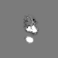
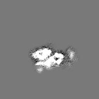
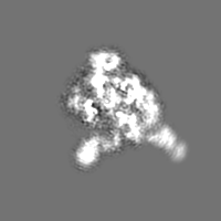
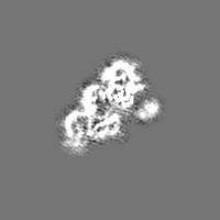
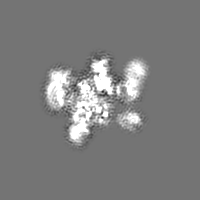
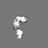
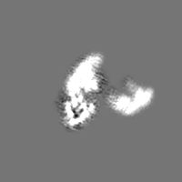
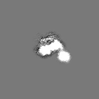
| Projections & slices | Image control
Images are generated by Spider. | ||||||||||||||||||||||||||||||||||||||||||||||||||||||||||||
| Voxel size | X=Y=Z: 1.30654 Å | ||||||||||||||||||||||||||||||||||||||||||||||||||||||||||||
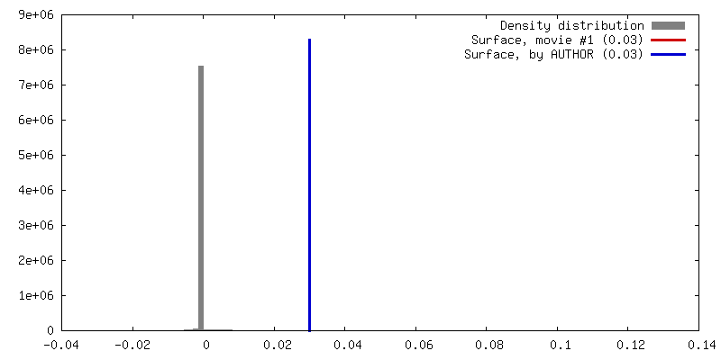
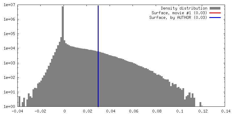
| Density |
| ||||||||||||||||||||||||||||||||||||||||||||||||||||||||||||
| Symmetry | Space group: 1 | ||||||||||||||||||||||||||||||||||||||||||||||||||||||||||||
| Details | EMDB XML:
CCP4 map header:
| ||||||||||||||||||||||||||||||||||||||||||||||||||||||||||||
 Z (Sec.)
 Y (Row.)
 X (Col.)


-Supplemental data
- Sample components
-Entire : Structure a of Group II Intron Complexed with its Reverse Transcr...
| Entire | Name: Structure a of Group II Intron Complexed with its Reverse Transcriptase |
|---|---|
| Components |
|
-Supramolecule #1000: Structure a of Group II Intron Complexed with its Reverse Transcr...
| Supramolecule | Name: Structure a of Group II Intron Complexed with its Reverse Transcriptase type: sample / ID: 1000 / Number unique components: 2 |
|---|---|
| Molecular weight | Experimental: 360 KDa / Theoretical: 360 KDa |
-Macromolecule #1: Group II Intron
| Macromolecule | Name: Group II Intron / type: rna / ID: 1 / Classification: OTHER / Structure: SINGLE STRANDED / Synthetic?: No |
|---|---|
| Source (natural) | Organism: Lactococcus lactis (lactic acid bacteria) |
-Macromolecule #2: Reverse Transcriptase
| Macromolecule | Name: Reverse Transcriptase / type: protein_or_peptide / ID: 2 / Recombinant expression: No |
|---|---|
| Source (natural) | Organism: Lactococcus lactis (lactic acid bacteria) |
-Experimental details
-Structure determination
| Method | cryo EM |
|---|---|
 Processing Processing | single particle reconstruction |
| Aggregation state | particle |
-Sample preparation
| Grid | Details: Quantifoil Cu-Rh R1.2/1.3 grid with thin carbon support |
|---|---|
| Vitrification | Cryogen name: ETHANE / Chamber humidity: 100 % / Instrument: FEI VITROBOT MARK IV |
- Electron microscopy
| Microscope | FEI TITAN KRIOS |
|---|---|
| Date | Oct 1, 2014 |
| Image recording | Category: CCD / Film or detector model: GATAN K2 SUMMIT (4k x 4k) / Number real images: 2266 |
| Electron beam | Acceleration voltage: 300 kV / Electron source:  FIELD EMISSION GUN FIELD EMISSION GUN |
| Electron optics | Illumination mode: FLOOD BEAM / Imaging mode: BRIGHT FIELD / Nominal defocus max: 3.0 µm / Nominal defocus min: 1.2 µm |
| Sample stage | Specimen holder model: FEI TITAN KRIOS AUTOGRID HOLDER |
| Experimental equipment |  Model: Titan Krios / Image courtesy: FEI Company |
-Image processing
| CTF correction | Details: each particle |
|---|---|
| Final reconstruction | Applied symmetry - Point group: C1 (asymmetric) / Resolution.type: BY AUTHOR / Resolution: 4.2 Å / Resolution method: OTHER / Software - Name: SPIDER, EMAN2, RELION / Number images used: 450296 |
