 Movie
Movie Controller


+ Open data

- Basic information
| Entry | 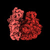 | |||||||||
|---|---|---|---|---|---|---|---|---|---|---|
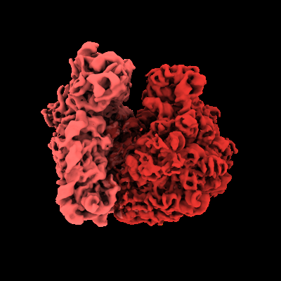
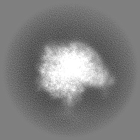
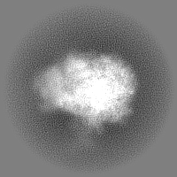
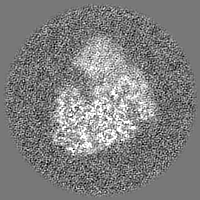
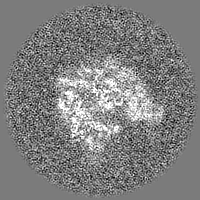
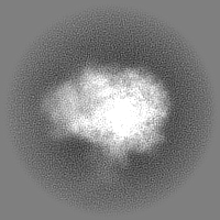
| Title | Subtomogram average of 70S ribosome (50S aligned) | |||||||||
 Map data Map data | ||||||||||
 Sample Sample |
| |||||||||
 Keywords Keywords | 70S / bacterial / RIBOSOME | |||||||||
| Biological species |  | |||||||||
| Method | subtomogram averaging / cryo EM / Resolution: 5.8 Å | |||||||||
 Authors Authors | Eisenstein F / Danev R | |||||||||
| Funding support |  Japan, 2 items Japan, 2 items
| |||||||||
 Citation Citation | Journal: Nat Methods / Year: 2023 Title: Parallel cryo electron tomography on in situ lamellae. Authors: Fabian Eisenstein / Haruaki Yanagisawa / Hiroka Kashihara / Masahide Kikkawa / Sachiko Tsukita / Radostin Danev / Abstract: In situ cryo electron tomography of cryo focused ion beam milled samples has emerged in recent years as a powerful technique for structural studies of macromolecular complexes in their native ...In situ cryo electron tomography of cryo focused ion beam milled samples has emerged in recent years as a powerful technique for structural studies of macromolecular complexes in their native cellular environment. However, the possibilities for recording tomographic tilt series in a high-throughput manner are limited, in part by the lamella-shaped samples. Here we utilize a geometrical sample model and optical image shift to record tens of tilt series in parallel, thereby saving time and gaining access to sample areas conventionally used for tracking specimen movement. The parallel cryo electron tomography (PACE-tomo) method achieves a throughput faster than 5 min per tilt series and allows for the collection of sample areas that were previously unreachable, thus maximizing the amount of data from each lamella. Performance testing with ribosomes in vitro and in situ on state-of-the-art and general-purpose microscopes demonstrated the high throughput and quality of PACE-tomo. #1: Journal: Biorxiv / Year: 2022Title: Parallel cryo electron tomography on in situ lamellae. Authors: Eisenstein F / Yanagisawa H / Kashihara H / Kikkawa M / Tsukita S / Danev R | |||||||||
| History |
|
- Structure visualization
| Supplemental images |
|---|
- Downloads & links
-EMDB archive
| Map data | emd_33116.map.gz | 17.8 MB |  EMDB map data format EMDB map data format | |
|---|---|---|---|---|
| Header (meta data) | emd-33116-v30.xmlemd-33116.xml | 16.9 KB 16.9 KB | Display Display | EMDB header |
| FSC (resolution estimation) | emd_33116_fsc.xmlemd_33116_fsc_2.xml | 7.1 KB 7.1 KB | Display Display | FSC data file |
| Images |  emd_33116.png emd_33116.png | 97.1 KB | ||
| Masks | emd_33116_msk_1.mapemd_33116_msk_2.map | 30.5 MB 30.5 MB | Mask map | |
| Filedesc metadata | emd-33116.cif.gz | 4.6 KB | ||
| Others | emd_33116_half_map_1.map.gzemd_33116_half_map_2.map.gz | 15.1 MB 15.1 MB | ||
| Archive directory |  http://ftp.pdbj.org/pub/emdb/structures/EMD-33116ftp://ftp.pdbj.org/pub/emdb/structures/EMD-33116 http://ftp.pdbj.org/pub/emdb/structures/EMD-33116ftp://ftp.pdbj.org/pub/emdb/structures/EMD-33116 | HTTPS FTP |
-Validation report
| Summary document | emd_33116_validation.pdf.gz | 165.1 KB | Display | EMDB validaton report |
|---|---|---|---|---|
| Full document | emd_33116_full_validation.pdf.gz | 164.7 KB | Display | |
| Data in XML | emd_33116_validation.xml.gz | 497 B | Display | |
| Data in CIF | emd_33116_validation.cif.gz | 373 B | Display | |
| Arichive directory | https://ftp.pdbj.org/pub/emdb/validation_reports/EMD-33116ftp://ftp.pdbj.org/pub/emdb/validation_reports/EMD-33116 | HTTPS FTP |
-Related structure data
| Related structure data | C: citing same article ( |
|---|---|
| EM raw data | EMPIAR-10986 (Title: Parallel cryo electron tomography (PACE-tomo) of 70S ribosomes (200 kV, side-entry holder) Data size: 222.8 Data #1: Movie frames for all tilt series gain corrected with outdated gain reference [micrographs - multiframe] Data #2: Tilt series and meta data [tilt series]) |


-Links
| EMDB pages | EMDB (EBI/PDBe) / EMDataResource |
|---|
-Map
| File | Download / File: emd_33116.map.gz / Format: CCP4 / Size: 30.5 MB / Type: IMAGE STORED AS FLOATING POINT NUMBER (4 BYTES) | ||||||||||||||||||||
|---|---|---|---|---|---|---|---|---|---|---|---|---|---|---|---|---|---|---|---|---|---|
| Voxel size | X=Y=Z: 2.16 Å | ||||||||||||||||||||
| Density |
| ||||||||||||||||||||
| Symmetry | Space group: 1 | ||||||||||||||||||||
| Details | EMDB XML:
|
-Supplemental data
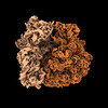
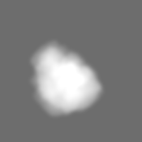
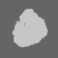
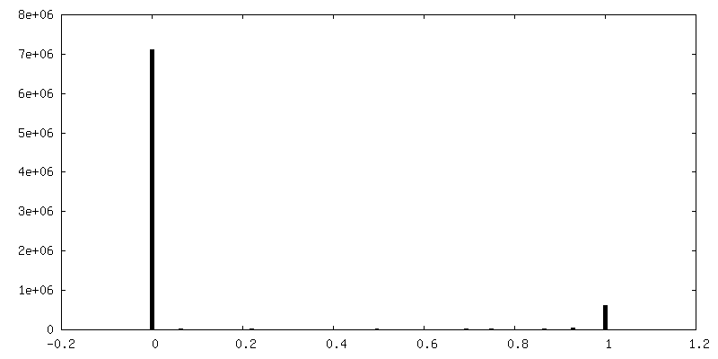
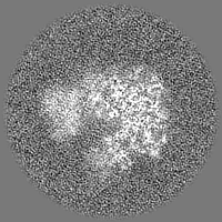
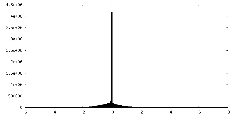
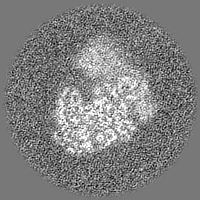
-Mask #1
| File | emd_33116_msk_1.map | ||||||||||||
|---|---|---|---|---|---|---|---|---|---|---|---|---|---|
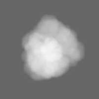
| Projections & Slices |
| ||||||||||||
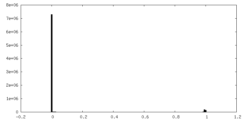
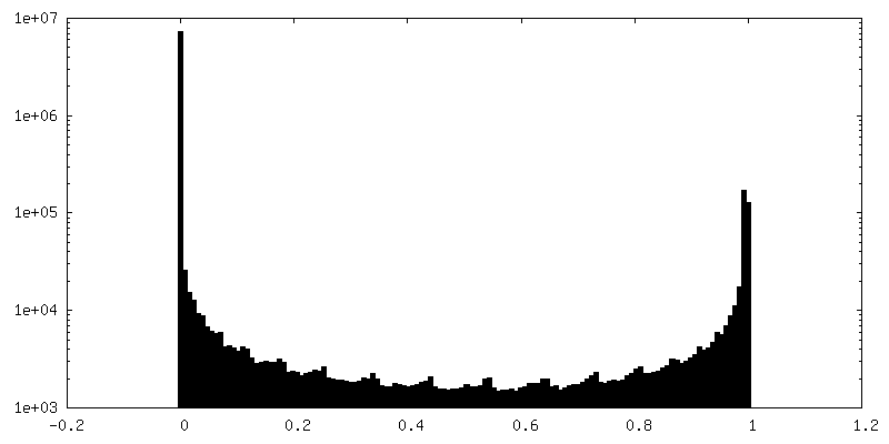
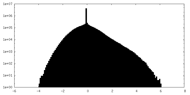
| Density Histograms |
 Z
Z Y
Y X
X
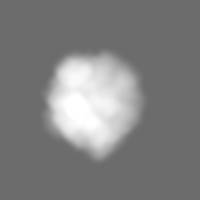


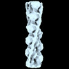
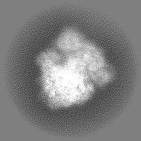
-Mask #2
| File | emd_33116_msk_2.map | ||||||||||||
|---|---|---|---|---|---|---|---|---|---|---|---|---|---|
| Projections & Slices |
| ||||||||||||
| Density Histograms |


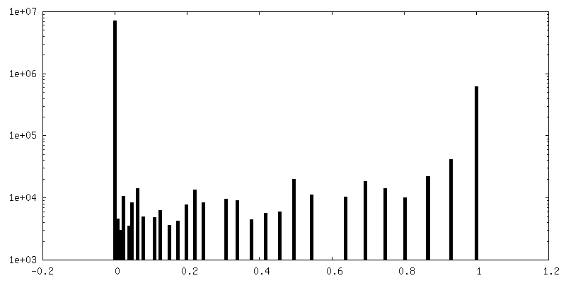
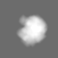
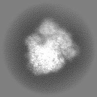
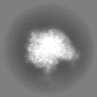
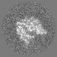
-Half map: #2
| File | emd_33116_half_map_1.map | ||||||||||||
|---|---|---|---|---|---|---|---|---|---|---|---|---|---|
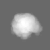
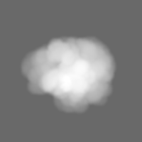
| Projections & Slices |
| ||||||||||||
| Density Histograms |

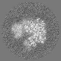
-Half map: #1
| File | emd_33116_half_map_2.map | ||||||||||||
|---|---|---|---|---|---|---|---|---|---|---|---|---|---|
| Projections & Slices |
| ||||||||||||
| Density Histograms |

- Sample components
-Entire : 70S ribosome (50S aligned)
| Entire | Name: 70S ribosome (50S aligned) |
|---|---|
| Components |
|
-Supramolecule #1: 70S ribosome (50S aligned)
| Supramolecule | Name: 70S ribosome (50S aligned) / type: complex / ID: 1 / Parent: 0 |
|---|---|
| Source (natural) | Organism: |
-Experimental details
-Structure determination
| Method | cryo EM |
|---|---|
 Processing Processing | subtomogram averaging |
| Aggregation state | particle |
-Sample preparation
| Concentration | 1.8 mg/mL | |||||||||||||||
|---|---|---|---|---|---|---|---|---|---|---|---|---|---|---|---|---|
| Buffer | pH: 7.5 Component:
| |||||||||||||||
| Grid | Model: Quantifoil R1.2/1.3 / Material: COPPER / Mesh: 200 / Support film - Material: CARBON / Support film - topology: HOLEY / Pretreatment - Type: GLOW DISCHARGE / Pretreatment - Time: 60 sec. / Pretreatment - Atmosphere: AIR | |||||||||||||||
| Vitrification | Cryogen name: ETHANE-PROPANE / Chamber humidity: 100 % / Chamber temperature: 277 K / Instrument: FEI VITROBOT MARK IV |
- Electron microscopy
| Microscope | JEOL 2010F |
|---|---|
| Details | Microscope was actually a JEOL JEM-F200 |
| Image recording | Film or detector model: GATAN K2 SUMMIT (4k x 4k) / Detector mode: COUNTING / Number grids imaged: 1 / Average exposure time: 2.1 sec. / Average electron dose: 5.4 e/Å2 Details: Tilt series were recorded in parallel using beam image shift |
| Electron beam | Acceleration voltage: 200 kV / Electron source:  FIELD EMISSION GUN FIELD EMISSION GUN |
| Electron optics | C2 aperture diameter: 40.0 µm / Illumination mode: FLOOD BEAM / Imaging mode: BRIGHT FIELD / Cs: 2.0 mm / Nominal defocus max: 4.0 µm / Nominal defocus min: 4.0 µm |
| Sample stage | Specimen holder model: GATAN 626 SINGLE TILT LIQUID NITROGEN CRYO TRANSFER HOLDER |
-Image processing
| Final reconstruction | Applied symmetry - Point group: C1 (asymmetric) / Resolution.type: BY AUTHOR / Resolution: 5.8 Å / Resolution method: FSC 0.143 CUT-OFF / Software - Name: RELION (ver. 4) / Details: 5.8 Angstrom for full 70S ribosome / Number subtomograms used: 10540 |
|---|---|
| Extraction | Number tomograms: 25 / Number images used: 53982 / Software - Name: crYOLO |
| Final angle assignment | Type: MAXIMUM LIKELIHOOD / Software - Name: RELION (ver. 4) |
| FSC plot (resolution estimation) | 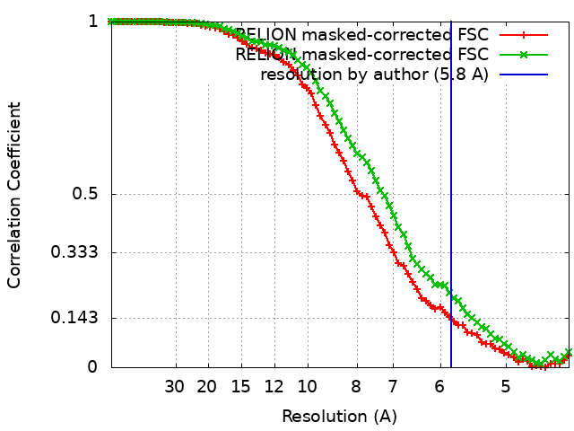 |
