 Movie
Movie Controller

[English] 日本語
 Yorodumi
- EMDB-33036: Local refinement map of Zur regions in Streptomyces coelicolor RN... -
+ Open data

- Basic information
| Entry |  | |||||||||
|---|---|---|---|---|---|---|---|---|---|---|
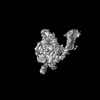
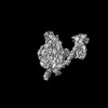
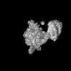
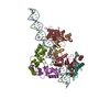
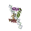
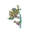
| Title | Local refinement map of Zur regions in Streptomyces coelicolor RNAP-promoter open complex with three Zur dimers | |||||||||
 Map data Map data | ||||||||||
 Sample Sample |
| |||||||||
| Biological species |  Streptomyces coelicolor A3(2) (bacteria) Streptomyces coelicolor A3(2) (bacteria) | |||||||||
| Method | single particle reconstruction / cryo EM / Resolution: 5.37 Å | |||||||||
 Authors Authors | Yang X / Zheng J | |||||||||
| Funding support |  China, 1 items China, 1 items
| |||||||||
 Citation Citation | Journal: Nucleic Acids Res / Year: 2022 Title: Structural basis of Streptomyces transcription activation by zinc uptake regulator. Authors: Xu Yang / Yiqun Wang / Guiyang Liu / Zixin Deng / Shuangjun Lin / Jianting Zheng / Abstract: Streptomyces coelicolor (Sc) is a model organism of actinobacteria to study morphological differentiation and production of bioactive metabolites. Sc zinc uptake regulator (Zur) affects both ...Streptomyces coelicolor (Sc) is a model organism of actinobacteria to study morphological differentiation and production of bioactive metabolites. Sc zinc uptake regulator (Zur) affects both processes by controlling zinc homeostasis. It activates transcription by binding to palindromic Zur-box sequences upstream of -35 elements. Here we deciphered the molecular mechanism by which ScZur interacts with promoter DNA and Sc RNA polymerase (RNAP) by cryo-EM structures and biochemical assays. The ScZur-DNA structures reveal a sequential and cooperative binding of three ScZur dimers surrounding a Zur-box spaced 8 nt upstream from a -35 element. The ScRNAPσHrdB-Zur-DNA structures define protein-protein and protein-DNA interactions involved in the principal housekeeping σHrdB-dependent transcription initiation from a noncanonical promoter with a -10 element lacking the critical adenine residue at position -11 and a TTGCCC -35 element deviating from the canonical TTGACA motif. ScZur interacts with the C-terminal domain of ScRNAP α subunit (αCTD) in a complex structure trapped in an active conformation. Key ScZur-αCTD interfacial residues accounting for ScZur-dependent transcription activation were confirmed by mutational studies. Together, our structural and biochemical results provide a comprehensive model for transcription activation of Zur family regulators. | |||||||||
| History |
|
- Structure visualization
| Supplemental images |
|---|

- Downloads & links
-EMDB archive
| Map data | emd_33036.map.gz | 203.6 MB |  EMDB map data format EMDB map data format | |
|---|---|---|---|---|
| Header (meta data) | emd-33036-v30.xmlemd-33036.xml | 15 KB 15 KB | Display Display | EMDB header |
| FSC (resolution estimation) | emd_33036_fsc.xml | 12.7 KB | Display | FSC data file |
| Images | 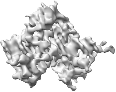 emd_33036.png emd_33036.png | 73.6 KB | ||
| Masks | emd_33036_msk_1.map | 216 MB | Mask map | |
| Others | emd_33036_half_map_1.map.gzemd_33036_half_map_2.map.gz | 200.6 MB 200.6 MB | ||
| Archive directory |  http://ftp.pdbj.org/pub/emdb/structures/EMD-33036ftp://ftp.pdbj.org/pub/emdb/structures/EMD-33036 http://ftp.pdbj.org/pub/emdb/structures/EMD-33036ftp://ftp.pdbj.org/pub/emdb/structures/EMD-33036 | HTTPS FTP |
-Validation report
| Summary document | emd_33036_validation.pdf.gz | 839.4 KB | Display | EMDB validaton report |
|---|---|---|---|---|
| Full document | emd_33036_full_validation.pdf.gz | 838.9 KB | Display | |
| Data in XML | emd_33036_validation.xml.gz | 21.2 KB | Display | |
| Data in CIF | emd_33036_validation.cif.gz | 27.4 KB | Display | |
| Arichive directory | https://ftp.pdbj.org/pub/emdb/validation_reports/EMD-33036ftp://ftp.pdbj.org/pub/emdb/validation_reports/EMD-33036 | HTTPS FTP |
-Related structure data


-Links
| EMDB pages | EMDB (EBI/PDBe) / EMDataResource |
|---|
-Map
| File | Download / File: emd_33036.map.gz / Format: CCP4 / Size: 216 MB / Type: IMAGE STORED AS FLOATING POINT NUMBER (4 BYTES) | ||||||||||||||||||||||||||||||||||||
|---|---|---|---|---|---|---|---|---|---|---|---|---|---|---|---|---|---|---|---|---|---|---|---|---|---|---|---|---|---|---|---|---|---|---|---|---|---|
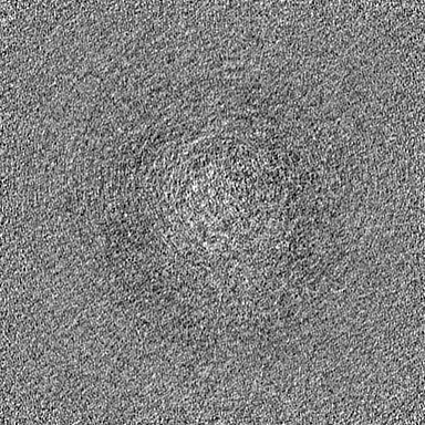
| Projections & slices | Image control
Images are generated by Spider. | ||||||||||||||||||||||||||||||||||||
| Voxel size | X=Y=Z: 1.05 Å | ||||||||||||||||||||||||||||||||||||
| Density |
| ||||||||||||||||||||||||||||||||||||
| Symmetry | Space group: 1 | ||||||||||||||||||||||||||||||||||||
| Details | EMDB XML:
|
 Z (Sec.)
 Y (Row.)
 X (Col.)


-Supplemental data
-Mask #1
| File | emd_33036_msk_1.map | ||||||||||||
|---|---|---|---|---|---|---|---|---|---|---|---|---|---|
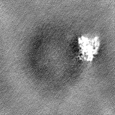
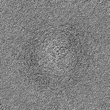
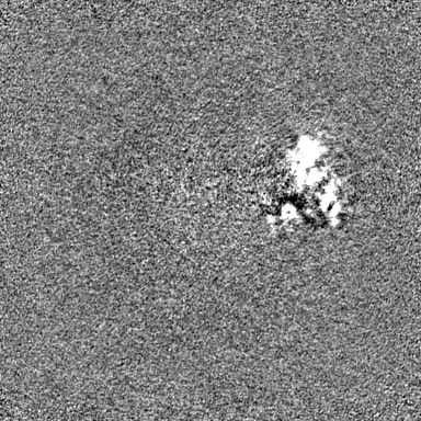
| Projections & Slices |
| ||||||||||||
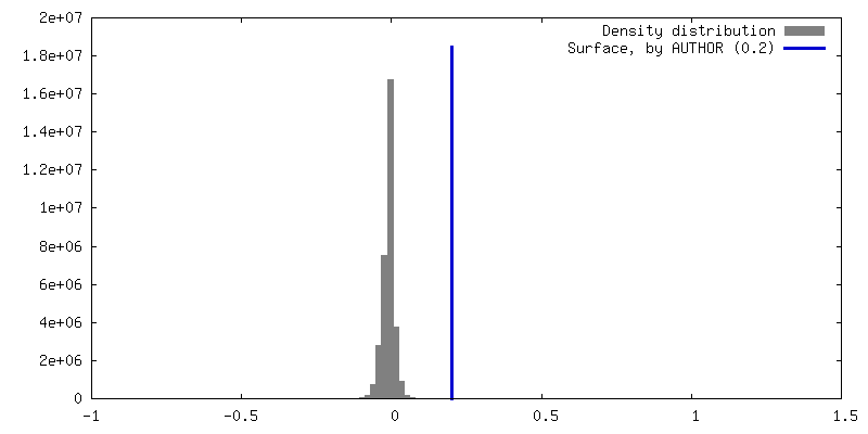
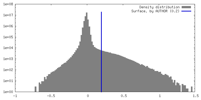
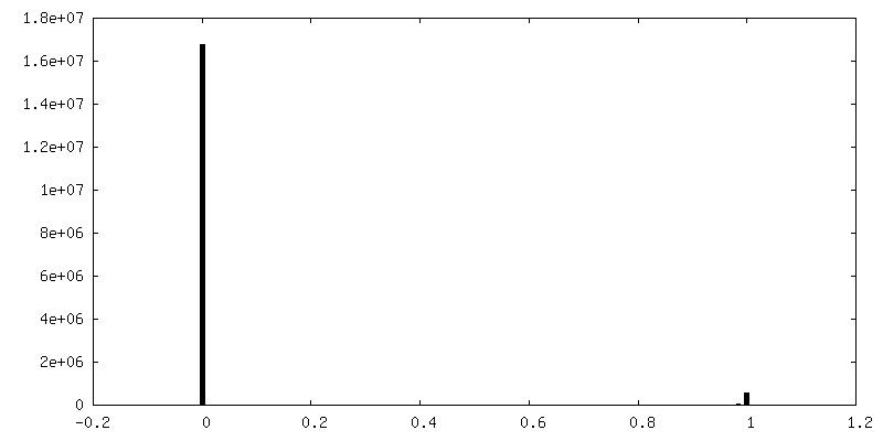
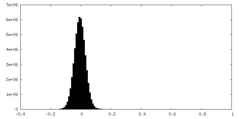
| Density Histograms |


-Half map: #2
| File | emd_33036_half_map_1.map | ||||||||||||
|---|---|---|---|---|---|---|---|---|---|---|---|---|---|
| Projections & Slices |
| ||||||||||||
| Density Histograms |


-Half map: #1
| File | emd_33036_half_map_2.map | ||||||||||||
|---|---|---|---|---|---|---|---|---|---|---|---|---|---|
| Projections & Slices |
| ||||||||||||
| Density Histograms |


- Sample components
-Entire : Local refinement map of Zur regions in Streptomyces coelicolor RN...
| Entire | Name: Local refinement map of Zur regions in Streptomyces coelicolor RNAP-promoter open complex with three Zur dimers |
|---|---|
| Components |
|
-Supramolecule #1: Local refinement map of Zur regions in Streptomyces coelicolor RN...
| Supramolecule | Name: Local refinement map of Zur regions in Streptomyces coelicolor RNAP-promoter open complex with three Zur dimers type: complex / Chimera: Yes / ID: 1 / Parent: 0 / Macromolecule list: #1-#9 |
|---|---|
| Source (natural) | Organism: Streptomyces coelicolor A3(2) (bacteria) |
| Recombinant expression | Organism: |
| Molecular weight | Experimental: 560 KDa |
-Experimental details
-Structure determination
| Method | cryo EM |
|---|---|
 Processing Processing | single particle reconstruction |
| Aggregation state | particle |
-Sample preparation
| Concentration | 0.8 mg/mL | ||||||||||||||||||
|---|---|---|---|---|---|---|---|---|---|---|---|---|---|---|---|---|---|---|---|
| Buffer | pH: 8 Component:
| ||||||||||||||||||
| Vitrification | Cryogen name: ETHANE / Chamber humidity: 100 % / Chamber temperature: 289 K / Instrument: FEI VITROBOT MARK IV |
- Electron microscopy
| Microscope | FEI TITAN KRIOS |
|---|---|
| Image recording | Film or detector model: GATAN K2 QUANTUM (4k x 4k) / Average electron dose: 1.25 e/Å2 |
| Electron beam | Acceleration voltage: 300 kV / Electron source:  FIELD EMISSION GUN FIELD EMISSION GUN |
| Electron optics | Illumination mode: FLOOD BEAM / Imaging mode: BRIGHT FIELD / Nominal defocus max: 2.0 µm / Nominal defocus min: 1.0 µm |
| Experimental equipment |  Model: Titan Krios / Image courtesy: FEI Company |
-Image processing
| Final reconstruction | Resolution.type: BY AUTHOR / Resolution: 5.37 Å / Resolution method: FSC 0.143 CUT-OFF / Number images used: 30218 |
|---|---|
| Initial angle assignment | Type: NOT APPLICABLE |
| Final angle assignment | Type: NOT APPLICABLE |
| FSC plot (resolution estimation) |  |
