 Movie
Movie Controller


+ Open data

- Basic information
| Entry | 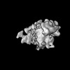 | |||||||||
|---|---|---|---|---|---|---|---|---|---|---|
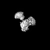
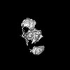
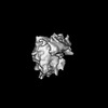
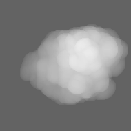
| Title | the neuron ribosome bound to the membrane | |||||||||
 Map data Map data | ||||||||||
 Sample Sample |
| |||||||||
| Biological species |  | |||||||||
| Method | subtomogram averaging / cryo EM / Resolution: 27.9 Å | |||||||||
 Authors Authors | Jiang W / Guo Q | |||||||||
| Funding support | 1 items
| |||||||||
 Citation Citation | Journal: Nucleic Acids Res / Year: 2022 Title: A transformation clustering algorithm and its application in polyribosomes structural profiling. Authors: Wenhong Jiang / Jonathan Wagner / Wenjing Du / Juergen Plitzko / Wolfgang Baumeister / Florian Beck / Qiang Guo /   Abstract: Improvements in cryo-electron tomography sample preparation, electron-microscopy instrumentations, and image processing algorithms have advanced the structural analysis of macromolecules in situ. ...Improvements in cryo-electron tomography sample preparation, electron-microscopy instrumentations, and image processing algorithms have advanced the structural analysis of macromolecules in situ. Beyond such analyses of individual macromolecules, the study of their interactions with functionally related neighbors in crowded cellular habitats, i.e. 'molecular sociology', is of fundamental importance in biology. Here we present a NEighboring Molecule TOpology Clustering (NEMO-TOC) algorithm. We optimized this algorithm for the detection and profiling of polyribosomes, which play both constitutive and regulatory roles in gene expression. Our results suggest a model where polysomes are formed by connecting multiple nonstochastic blocks, in which translation is likely synchronized. | |||||||||
| History |
|
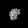
- Structure visualization
| Supplemental images |
|---|

- Downloads & links
-EMDB archive
| Map data | emd_33018.map.gz | 28 MB |  EMDB map data format EMDB map data format | |
|---|---|---|---|---|
| Header (meta data) | emd-33018-v30.xmlemd-33018.xml | 11.6 KB 11.6 KB | Display Display | EMDB header |
| Images |  emd_33018.png emd_33018.png | 31.6 KB | ||
| Masks | emd_33018_msk_1.map | 26.2 MB | Mask map | |
| Others | emd_33018_half_map_1.map.gzemd_33018_half_map_2.map.gz | 16.6 MB 16.6 MB | ||
| Archive directory |  http://ftp.pdbj.org/pub/emdb/structures/EMD-33018ftp://ftp.pdbj.org/pub/emdb/structures/EMD-33018 http://ftp.pdbj.org/pub/emdb/structures/EMD-33018ftp://ftp.pdbj.org/pub/emdb/structures/EMD-33018 | HTTPS FTP |
-Validation report
| Summary document | emd_33018_validation.pdf.gz | 768.4 KB | Display | EMDB validaton report |
|---|---|---|---|---|
| Full document | emd_33018_full_validation.pdf.gz | 768 KB | Display | |
| Data in XML | emd_33018_validation.xml.gz | 10.9 KB | Display | |
| Data in CIF | emd_33018_validation.cif.gz | 12.6 KB | Display | |
| Arichive directory | https://ftp.pdbj.org/pub/emdb/validation_reports/EMD-33018ftp://ftp.pdbj.org/pub/emdb/validation_reports/EMD-33018 | HTTPS FTP |
-Related structure data
-Links
| EMDB pages | EMDB (EBI/PDBe) / EMDataResource |
|---|
-Map
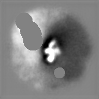
| File | Download / File: emd_33018.map.gz / Format: CCP4 / Size: 32.4 MB / Type: IMAGE STORED AS FLOATING POINT NUMBER (4 BYTES) | ||||||||||||||||||||||||||||||||||||
|---|---|---|---|---|---|---|---|---|---|---|---|---|---|---|---|---|---|---|---|---|---|---|---|---|---|---|---|---|---|---|---|---|---|---|---|---|---|
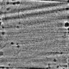
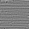
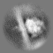
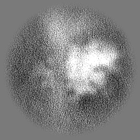
| Projections & slices | Image control
Images are generated by Spider. | ||||||||||||||||||||||||||||||||||||
| Voxel size | X=Y=Z: 3.42 Å | ||||||||||||||||||||||||||||||||||||
| Density |
| ||||||||||||||||||||||||||||||||||||
| Symmetry | Space group: 1 | ||||||||||||||||||||||||||||||||||||
| Details | EMDB XML:
|
 Z (Sec.)
 Y (Row.)
 X (Col.)


-Supplemental data
-Mask #1
| File | emd_33018_msk_1.map | ||||||||||||
|---|---|---|---|---|---|---|---|---|---|---|---|---|---|
| Projections & Slices |
| ||||||||||||
| Density Histograms |


-Half map: #2
| File | emd_33018_half_map_1.map | ||||||||||||
|---|---|---|---|---|---|---|---|---|---|---|---|---|---|
| Projections & Slices |
| ||||||||||||
| Density Histograms |


-Half map: #1
| File | emd_33018_half_map_2.map | ||||||||||||
|---|---|---|---|---|---|---|---|---|---|---|---|---|---|
| Projections & Slices |
| ||||||||||||
| Density Histograms |


- Sample components
-Entire : ribosome bound to the ER membrane
| Entire | Name: ribosome bound to the ER membrane |
|---|---|
| Components |
|
-Supramolecule #1: ribosome bound to the ER membrane
| Supramolecule | Name: ribosome bound to the ER membrane / type: complex / Chimera: Yes / ID: 1 / Parent: 0 |
|---|---|
| Source (natural) | Organism: |
-Experimental details
-Structure determination
| Method | cryo EM |
|---|---|
 Processing Processing | subtomogram averaging |
| Aggregation state | cell |
-Sample preparation
| Buffer | pH: 7 |
|---|---|
| Vitrification | Cryogen name: ETHANE |
- Electron microscopy
| Microscope | FEI TITAN KRIOS |
|---|---|
| Image recording | Film or detector model: GATAN K2 SUMMIT (4k x 4k) / Average electron dose: 1.8 e/Å2 |
| Electron beam | Acceleration voltage: 300 kV / Electron source:  FIELD EMISSION GUN FIELD EMISSION GUN |
| Electron optics | Illumination mode: OTHER / Imaging mode: BRIGHT FIELD / Nominal defocus max: 6.0 µm / Nominal defocus min: 3.0 µm |
| Experimental equipment |  Model: Titan Krios / Image courtesy: FEI Company |
-Image processing
| Final reconstruction | Resolution.type: BY AUTHOR / Resolution: 27.9 Å / Resolution method: FSC 0.143 CUT-OFF / Number subtomograms used: 1166 |
|---|---|
| Extraction | Number tomograms: 18 / Number images used: 11547 |
| Final angle assignment | Type: MAXIMUM LIKELIHOOD |
