 Movie
Movie Controller


+ Open data

- Basic information
| Entry |  | |||||||||
|---|---|---|---|---|---|---|---|---|---|---|
| Title | Overall structure of Helicobacter pylori bacteriophage KHP30 | |||||||||
 Map data Map data | ||||||||||
 Sample Sample |
| |||||||||
| Biological species |  Helicobacter phage KHP (virus) Helicobacter phage KHP (virus) | |||||||||
| Method | single particle reconstruction / cryo EM / Resolution: 6.4 Å | |||||||||
 Authors Authors | Kamiya R / Uchiyama J / Matsuzaki S / Murata K / Iwasaki K / Miyazaki N | |||||||||
| Funding support |  Japan, 2 items Japan, 2 items
| |||||||||
 Citation Citation | Journal: To Be Published Title: Tail structure of Helicobacter pylori bacteriophage KHP30 by single particle cryo-electron microscopy Authors: Kamiya R / Uchiyama J / Matsuzaki S / Murata K / Iwasaki K / Miyazaki N | |||||||||
| History |
|
- Structure visualization
| Supplemental images |
|---|

- Downloads & links
-EMDB archive
| Map data | emd_32614.map.gz | 34.4 MB |  EMDB map data format EMDB map data format | |
|---|---|---|---|---|
| Header (meta data) | emd-32614-v30.xmlemd-32614.xml | 8.9 KB 8.9 KB | Display Display | EMDB header |
| FSC (resolution estimation) | emd_32614_fsc.xml | 12.4 KB | Display | FSC data file |
| Images | 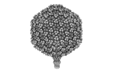 emd_32614.png emd_32614.png | 57 KB | ||
| Archive directory |  http://ftp.pdbj.org/pub/emdb/structures/EMD-32614ftp://ftp.pdbj.org/pub/emdb/structures/EMD-32614 http://ftp.pdbj.org/pub/emdb/structures/EMD-32614ftp://ftp.pdbj.org/pub/emdb/structures/EMD-32614 | HTTPS FTP |
-Related structure data
| Related structure data |
|---|

-Links
| EMDB pages | EMDB (EBI/PDBe) / EMDataResource |
|---|
-Map
| File | Download / File: emd_32614.map.gz / Format: CCP4 / Size: 163.6 MB / Type: IMAGE STORED AS FLOATING POINT NUMBER (4 BYTES) | ||||||||||||||||||||||||||||||||||||
|---|---|---|---|---|---|---|---|---|---|---|---|---|---|---|---|---|---|---|---|---|---|---|---|---|---|---|---|---|---|---|---|---|---|---|---|---|---|
| Projections & slices | Image control
Images are generated by Spider. | ||||||||||||||||||||||||||||||||||||
| Voxel size | X=Y=Z: 3.18 Å | ||||||||||||||||||||||||||||||||||||
| Density |
| ||||||||||||||||||||||||||||||||||||
| Symmetry | Space group: 1 | ||||||||||||||||||||||||||||||||||||
| Details | EMDB XML:
|
 Z (Sec.)
 Y (Row.)
 X (Col.)


-Supplemental data
- Sample components
-Entire : Helicobacter phage KHP
| Entire | Name: Helicobacter phage KHP (virus) |
|---|---|
| Components |
|
-Supramolecule #1: Helicobacter phage KHP
| Supramolecule | Name: Helicobacter phage KHP / type: virus / ID: 1 / Parent: 0 / Macromolecule list: #1-#3 / NCBI-ID: 1208236 / Sci species name: Helicobacter phage KHP / Virus type: VIRION / Virus isolate: OTHER / Virus enveloped: No / Virus empty: No |
|---|---|
| Virus shell | Shell ID: 1 / Name: Head / Diameter: 700.0 Å / T number (triangulation number): 9 |
-Experimental details
-Structure determination
| Method | cryo EM |
|---|---|
 Processing Processing | single particle reconstruction |
| Aggregation state | particle |
-Sample preparation
| Buffer | pH: 7.2 |
|---|---|
| Vitrification | Cryogen name: ETHANE / Chamber humidity: 100 % / Chamber temperature: 277 K / Instrument: FEI VITROBOT MARK IV |
- Electron microscopy
| Microscope | FEI TITAN KRIOS |
|---|---|
| Image recording | Film or detector model: FEI FALCON III (4k x 4k) / Detector mode: INTEGRATING / Average exposure time: 2.5 sec. / Average electron dose: 50.0 e/Å2 |
| Electron beam | Acceleration voltage: 300 kV / Electron source:  FIELD EMISSION GUN FIELD EMISSION GUN |
| Electron optics | Illumination mode: FLOOD BEAM / Imaging mode: BRIGHT FIELD / Cs: 2.7 mm / Nominal defocus max: 2.5 µm / Nominal defocus min: 1.0 µm |
| Sample stage | Specimen holder model: FEI TITAN KRIOS AUTOGRID HOLDER / Cooling holder cryogen: NITROGEN |
| Experimental equipment |  Model: Titan Krios / Image courtesy: FEI Company |

