Movie
Movie Controller

[English] 日本語
 Yorodumi
- EMDB-32611: Structure of Coxsackievirus A10 with Hybrid electron counting at ... -
+ Open data

- Basic information
| Entry |  | |||||||||
|---|---|---|---|---|---|---|---|---|---|---|
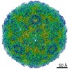
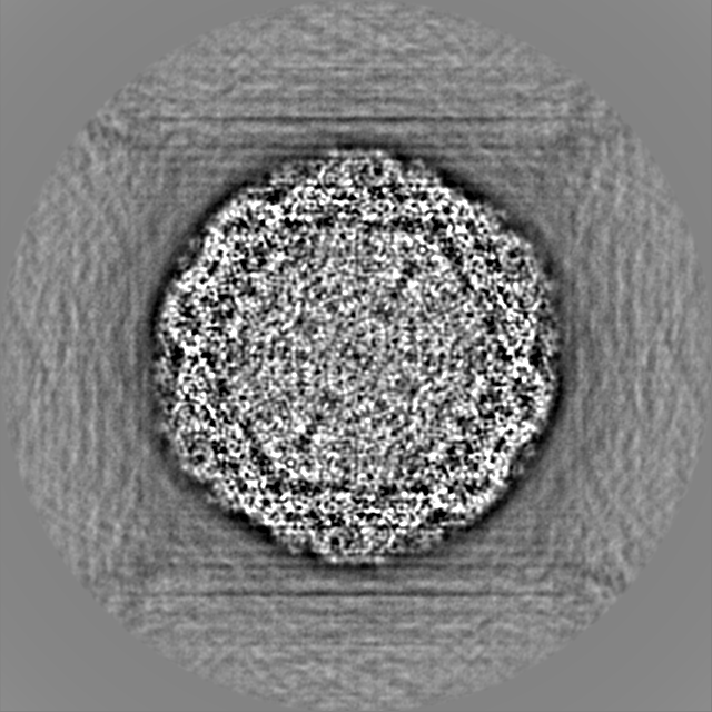
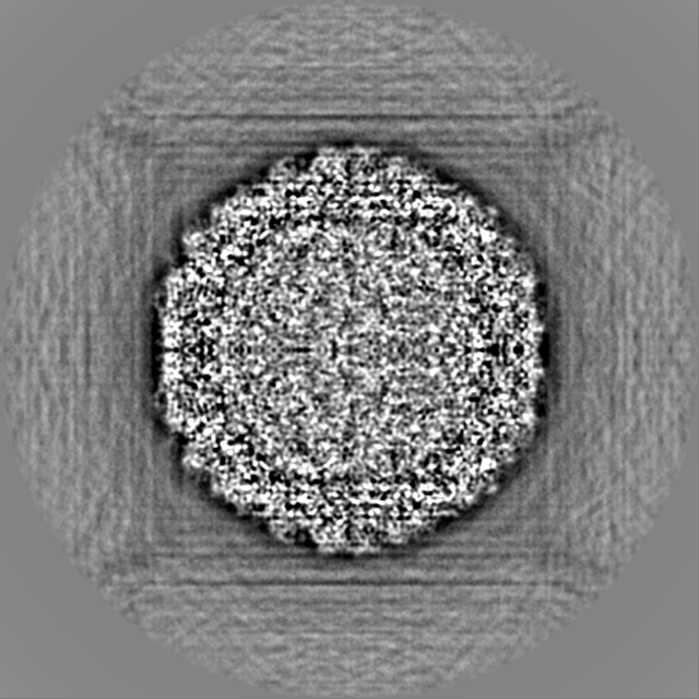
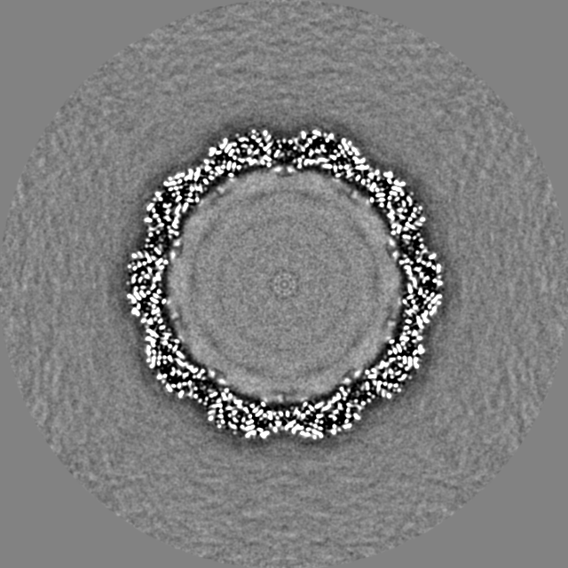
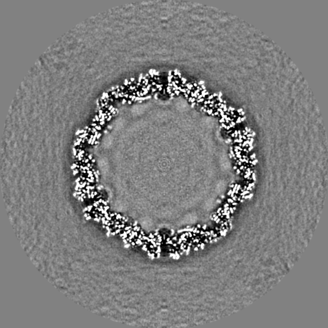
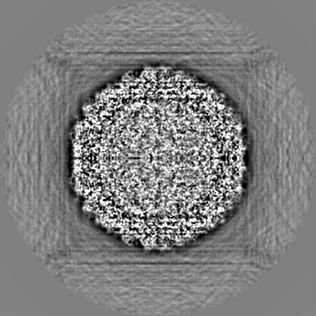
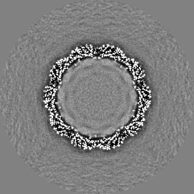
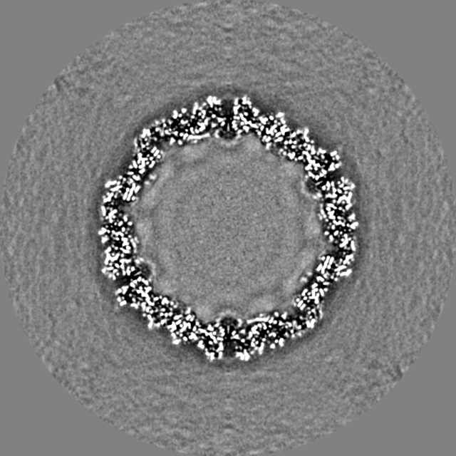
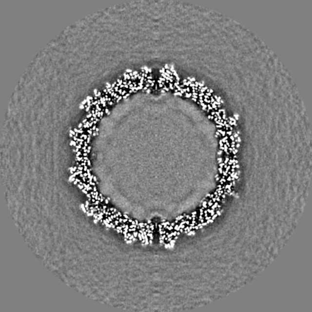
| Title | Structure of Coxsackievirus A10 with Hybrid electron counting at 200 kV | |||||||||
 Map data Map data | Coxsackievirus A10 Hybrid at 200 kV | |||||||||
 Sample Sample |
| |||||||||
 Keywords Keywords | Coxsackievirus A10 / VIRUS | |||||||||
| Biological species |  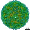 Coxsackievirus A10 Coxsackievirus A10 | |||||||||
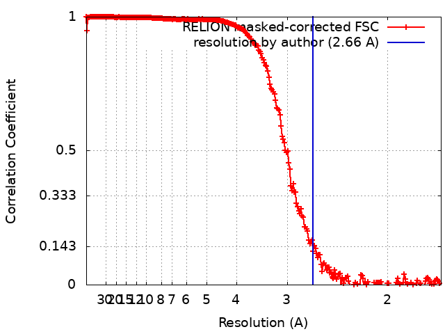
| Method | single particle reconstruction / cryo EM / Resolution: 2.66 Å | |||||||||
 Authors Authors | Zhu DJ / Zhang XZ | |||||||||
| Funding support |  China, 1 items China, 1 items
| |||||||||
 Citation Citation | Journal: Commun Biol / Year: 2022 Title: An electron counting algorithm improves imaging of proteins with low-acceleration-voltage cryo-electron microscope. Authors: Dongjie Zhu / Huigang Shi / Chunling Wu / Xinzheng Zhang / Abstract: Relative to the 300-kV accelerating field, electrons accelerated under lower voltages are potentially scattered more strongly. Lowering the accelerate voltage has been suggested to enhance the signal- ...Relative to the 300-kV accelerating field, electrons accelerated under lower voltages are potentially scattered more strongly. Lowering the accelerate voltage has been suggested to enhance the signal-to-noise ratio (SNR) of cryo-electron microscopy (cryo-EM) images of small-molecular-weight proteins (<100 kD). However, the detection efficient of current Direct Detection Devices (DDDs) and temporal coherence of cryo-EM decrease at lower voltage, leading to loss of SNR. Here, we present an electron counting algorithm to improve the detection of low-energy electrons. The counting algorithm increased the SNR of 120-kV and 200-kV cryo-EM image from a Falcon III camera by 8%, 20% at half the Nyquist frequency and 21%, 80% at Nyquist frequency, respectively, resulting in a considerable improvement in resolution of 3D reconstructions. Our results indicate that with further improved temporal coherence and a dedicated designed camera, a 120-kV cryo-electron microscope has potential to match the 300-kV microscope at imaging small proteins. | |||||||||
| History |
|
- Structure visualization
| Supplemental images |
|---|

- Downloads & links
-EMDB archive
| Map data | emd_32611.map.gz | 939.1 MB |  EMDB map data format EMDB map data format | |
|---|---|---|---|---|
| Header (meta data) | emd-32611-v30.xmlemd-32611.xml | 12.7 KB 12.7 KB | Display Display | EMDB header |
| FSC (resolution estimation) | emd_32611_fsc.xml | 22.5 KB | Display | FSC data file |
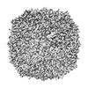
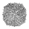
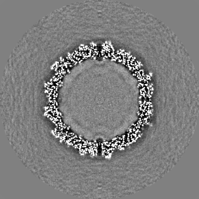
| Images | 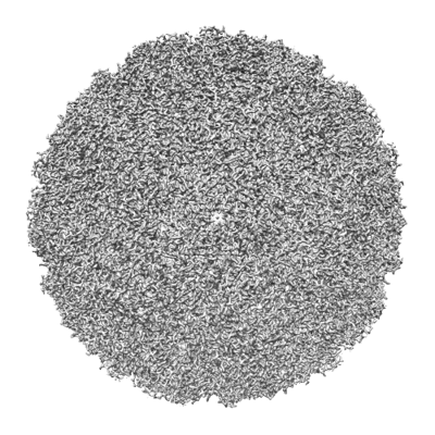 emd_32611.png emd_32611.png | 189.8 KB | ||
| Masks | emd_32611_msk_1.map | 1000 MB | Mask map | |
| Filedesc metadata | emd-32611.cif.gz | 3.8 KB | ||
| Others | emd_32611_half_map_1.map.gzemd_32611_half_map_2.map.gz | 816.6 MB 817 MB | ||
| Archive directory |  http://ftp.pdbj.org/pub/emdb/structures/EMD-32611ftp://ftp.pdbj.org/pub/emdb/structures/EMD-32611 http://ftp.pdbj.org/pub/emdb/structures/EMD-32611ftp://ftp.pdbj.org/pub/emdb/structures/EMD-32611 | HTTPS FTP |
-Validation report
| Summary document | emd_32611_validation.pdf.gz | 1.1 MB | Display | EMDB validaton report |
|---|---|---|---|---|
| Full document | emd_32611_full_validation.pdf.gz | 1.1 MB | Display | |
| Data in XML | emd_32611_validation.xml.gz | 30.6 KB | Display | |
| Data in CIF | emd_32611_validation.cif.gz | 40.9 KB | Display | |
| Arichive directory | https://ftp.pdbj.org/pub/emdb/validation_reports/EMD-32611ftp://ftp.pdbj.org/pub/emdb/validation_reports/EMD-32611 | HTTPS FTP |
-Related structure data
| Related structure data | C: citing same article ( |
|---|
-Links
| EMDB pages | EMDB (EBI/PDBe) / EMDataResource |
|---|
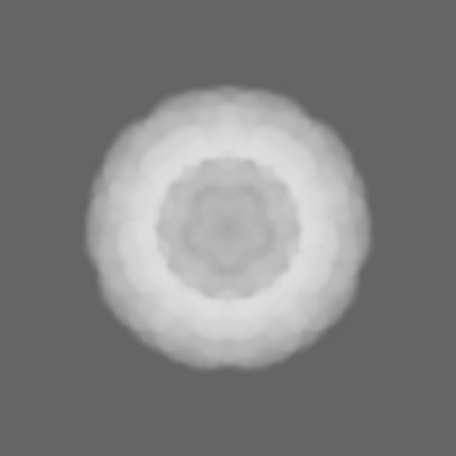
-Map
| File | Download / File: emd_32611.map.gz / Format: CCP4 / Size: 1000 MB / Type: IMAGE STORED AS FLOATING POINT NUMBER (4 BYTES) | ||||||||||||||||||||
|---|---|---|---|---|---|---|---|---|---|---|---|---|---|---|---|---|---|---|---|---|---|
| Annotation | Coxsackievirus A10 Hybrid at 200 kV | ||||||||||||||||||||
| Voxel size | X=Y=Z: 0.85 Å | ||||||||||||||||||||
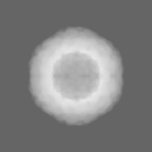
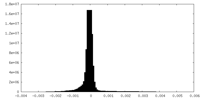
| Density |
| ||||||||||||||||||||
| Symmetry | Space group: 1 | ||||||||||||||||||||
| Details | EMDB XML:
|
-Supplemental data
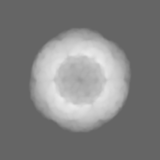
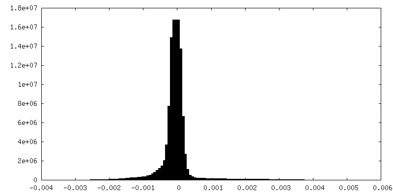
-Mask #1
| File | emd_32611_msk_1.map | ||||||||||||
|---|---|---|---|---|---|---|---|---|---|---|---|---|---|
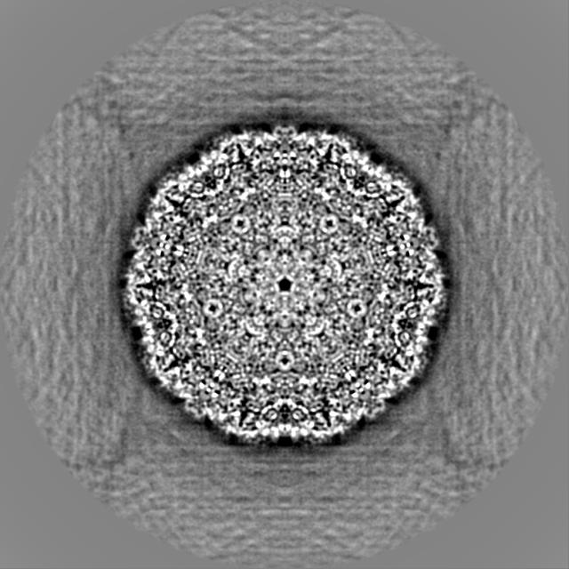
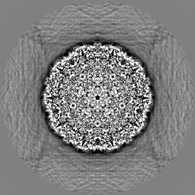
| Projections & Slices |
| ||||||||||||
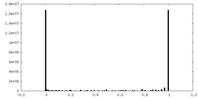
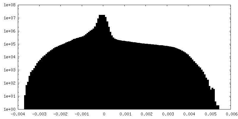
| Density Histograms |
 Z
Z Y
Y X
X


-Half map: Half2
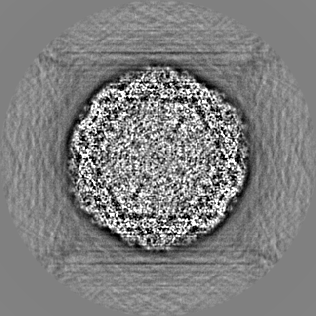
| File | emd_32611_half_map_1.map | ||||||||||||
|---|---|---|---|---|---|---|---|---|---|---|---|---|---|
| Annotation | Half2 | ||||||||||||
| Projections & Slices |
| ||||||||||||
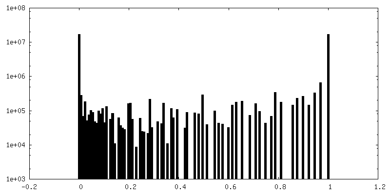
| Density Histograms |

-Half map: Half1
| File | emd_32611_half_map_2.map | ||||||||||||
|---|---|---|---|---|---|---|---|---|---|---|---|---|---|
| Annotation | Half1 | ||||||||||||
| Projections & Slices |
| ||||||||||||
| Density Histograms |
- Sample components
-Entire : Coxsackievirus A10
| Entire | Name: Coxsackievirus A10 |
|---|---|
| Components |
|
-Supramolecule #1: Coxsackievirus A10
| Supramolecule | Name: Coxsackievirus A10 / type: virus / ID: 1 / Parent: 0 / NCBI-ID: 42769 / Sci species name: Coxsackievirus A10 / Virus type: VIRION / Virus isolate: OTHER / Virus enveloped: No / Virus empty: No |
|---|
-Experimental details
-Structure determination
| Method | cryo EM |
|---|---|
 Processing Processing | single particle reconstruction |
| Aggregation state | particle |
-Sample preparation
| Buffer | pH: 7.4 |
|---|---|
| Vitrification | Cryogen name: ETHANE |
- Electron microscopy
| Microscope | FEI TITAN KRIOS |
|---|---|
| Image recording | Film or detector model: FEI FALCON III (4k x 4k) / Detector mode: OTHER / Digitization - Dimensions - Width: 4096 pixel / Digitization - Dimensions - Height: 4096 pixel / Average electron dose: 10.1 e/Å2 |
| Electron beam | Acceleration voltage: 200 kV / Electron source:  FIELD EMISSION GUN FIELD EMISSION GUN |
| Electron optics | Illumination mode: FLOOD BEAM / Imaging mode: BRIGHT FIELD / Nominal defocus max: 2.5 µm / Nominal defocus min: 0.6 µm |
| Experimental equipment |  Model: Titan Krios / Image courtesy: FEI Company |