 Movie
Movie Controller

[English] 日本語
 Yorodumi
- EMDB-32035: Structure of PA-tag modified E. hirae V-ATPase by Volta phase plate -
+ Open data

- Basic information
| Entry |  | |||||||||
|---|---|---|---|---|---|---|---|---|---|---|
| Title | Structure of PA-tag modified E. hirae V-ATPase by Volta phase plate | |||||||||
 Map data Map data | E. hirae V-ATPase with PA-tag | |||||||||
 Sample Sample |
| |||||||||
| Biological species |  Enterococcus hirae (bacteria) Enterococcus hirae (bacteria) | |||||||||
| Method | single particle reconstruction / cryo EM / Resolution: 6.7 Å | |||||||||
 Authors Authors | Burton-Smith RN / Murata K | |||||||||
| Funding support |  Japan, 1 items Japan, 1 items
| |||||||||
 Citation Citation | Journal: To Be Published Title: Examining the structure of E. hirae V-ATPase using Volta- and Zernike-type phase plate cryo-EM Authors: Burton-Smith RN / Murata K | |||||||||
| History |
|
- Structure visualization
| Supplemental images |
|---|

- Downloads & links
-EMDB archive
| Map data | emd_32035.map.gz | 113.3 MB |  EMDB map data format EMDB map data format | |
|---|---|---|---|---|
| Header (meta data) | emd-32035-v30.xmlemd-32035.xml | 7.1 KB 7.1 KB | Display Display | EMDB header |
| Images | 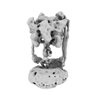 emd_32035.png emd_32035.png | 48.6 KB | ||
| Archive directory |  http://ftp.pdbj.org/pub/emdb/structures/EMD-32035ftp://ftp.pdbj.org/pub/emdb/structures/EMD-32035 http://ftp.pdbj.org/pub/emdb/structures/EMD-32035ftp://ftp.pdbj.org/pub/emdb/structures/EMD-32035 | HTTPS FTP |
-Links
| EMDB pages | EMDB (EBI/PDBe) / EMDataResource |
|---|
-Map
| File | Download / File: emd_32035.map.gz / Format: CCP4 / Size: 125 MB / Type: IMAGE STORED AS FLOATING POINT NUMBER (4 BYTES) | ||||||||||||||||||||||||||||||||||||
|---|---|---|---|---|---|---|---|---|---|---|---|---|---|---|---|---|---|---|---|---|---|---|---|---|---|---|---|---|---|---|---|---|---|---|---|---|---|
| Annotation | E. hirae V-ATPase with PA-tag | ||||||||||||||||||||||||||||||||||||
| Projections & slices | Image control
Images are generated by Spider. | ||||||||||||||||||||||||||||||||||||
| Voxel size | X=Y=Z: 1.113 Å | ||||||||||||||||||||||||||||||||||||
| Density |
| ||||||||||||||||||||||||||||||||||||
| Symmetry | Space group: 1 | ||||||||||||||||||||||||||||||||||||
| Details | EMDB XML:
|
 Z (Sec.)
 Y (Row.)
 X (Col.)


-Supplemental data
- Sample components
-Entire : V-ATPase with PA-tag fused to rotor
| Entire | Name: V-ATPase with PA-tag fused to rotor |
|---|---|
| Components |
|
-Supramolecule #1: V-ATPase with PA-tag fused to rotor
| Supramolecule | Name: V-ATPase with PA-tag fused to rotor / type: complex / ID: 1 / Parent: 0 |
|---|---|
| Source (natural) | Organism: Enterococcus hirae (bacteria) |
| Recombinant expression | Organism: |
-Experimental details
-Structure determination
| Method | cryo EM |
|---|---|
 Processing Processing | single particle reconstruction |
| Aggregation state | particle |
-Sample preparation
| Buffer | pH: 7.5 |
|---|---|
| Vitrification | Cryogen name: ETHANE |
- Electron microscopy
| Microscope | FEI TITAN KRIOS |
|---|---|
| Specialist optics | Phase plate: VOLTA PHASE PLATE |
| Image recording | Film or detector model: FEI FALCON II (4k x 4k) / Average electron dose: 54.0 e/Å2 |
| Electron beam | Acceleration voltage: 300 kV / Electron source:  FIELD EMISSION GUN FIELD EMISSION GUN |
| Electron optics | Illumination mode: FLOOD BEAM / Imaging mode: BRIGHT FIELD |
| Experimental equipment |  Model: Titan Krios / Image courtesy: FEI Company |
-Image processing
| Startup model | Type of model: OTHER / Details: Ab initio |
|---|---|
| Final reconstruction | Resolution.type: BY AUTHOR / Resolution: 6.7 Å / Resolution method: FSC 0.143 CUT-OFF / Number images used: 46959 |
| Initial angle assignment | Type: MAXIMUM LIKELIHOOD |
| Final angle assignment | Type: MAXIMUM LIKELIHOOD |
