 Movie
Movie Controller

[English] 日本語
 Yorodumi
- EMDB-31819: Negative staining (NS)-EM structure of SARS-CoV-2 S-Delta variant... -
+ Open data

- Basic information
| Entry |  | |||||||||||||||
|---|---|---|---|---|---|---|---|---|---|---|---|---|---|---|---|---|
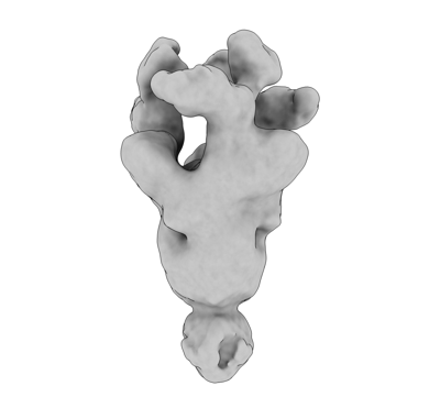
| Title | Negative staining (NS)-EM structure of SARS-CoV-2 S-Delta variant in complex with neutralizing antibodies, RBD-chAb-15 and RBD-chAb45 | |||||||||||||||
 Map data Map data | NSEM structure of SARS-CoV-2 S-Delta variant in complex with neutralizing antibodies, RBD-chAb-15 and RBD-chAb45 | |||||||||||||||
 Sample Sample |
| |||||||||||||||
| Biological species |   Severe acute respiratory syndrome coronavirus 2 Severe acute respiratory syndrome coronavirus 2 | |||||||||||||||
| Method | single particle reconstruction / negative staining / Resolution: 7.0 Å | |||||||||||||||
 Authors Authors | Yu PY / Yang TJ / Chang YC / Wu HC / Hsu STD | |||||||||||||||
| Funding support |  Taiwan, 4 items Taiwan, 4 items
| |||||||||||||||
 Citation Citation | Journal: To Be Published Title: Negative staining (NS)-EM structure of SARS-CoV-2 S-Delta variant in complex with neutralizing antibodies, RBD-chAb-15 and RBD-chAb45 Authors: Yu PY / Yang TJ / Chang YC / Wu HC / Hsu STD | |||||||||||||||
| History |
|
- Structure visualization
| Supplemental images |
|---|
- Downloads & links
-EMDB archive
| Map data | emd_31819.map.gz | 31.5 MB |  EMDB map data format EMDB map data format | |
|---|---|---|---|---|
| Header (meta data) | emd-31819-v30.xmlemd-31819.xml | 10.9 KB 10.9 KB | Display Display | EMDB header |
| Images |  emd_31819.png emd_31819.png | 34.5 KB | ||
| Archive directory |  http://ftp.pdbj.org/pub/emdb/structures/EMD-31819ftp://ftp.pdbj.org/pub/emdb/structures/EMD-31819 http://ftp.pdbj.org/pub/emdb/structures/EMD-31819ftp://ftp.pdbj.org/pub/emdb/structures/EMD-31819 | HTTPS FTP |
-Links
| EMDB pages | EMDB (EBI/PDBe) / EMDataResource |
|---|
-Map
| File | Download / File: emd_31819.map.gz / Format: CCP4 / Size: 64 MB / Type: IMAGE STORED AS FLOATING POINT NUMBER (4 BYTES) | ||||||||||||||||||||||||||||||||||||
|---|---|---|---|---|---|---|---|---|---|---|---|---|---|---|---|---|---|---|---|---|---|---|---|---|---|---|---|---|---|---|---|---|---|---|---|---|---|
| Annotation | NSEM structure of SARS-CoV-2 S-Delta variant in complex with neutralizing antibodies, RBD-chAb-15 and RBD-chAb45 | ||||||||||||||||||||||||||||||||||||
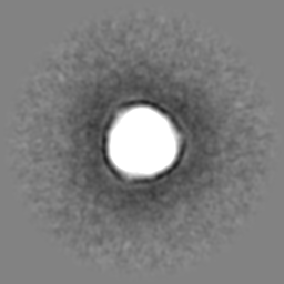
| Projections & slices | Image control
Images are generated by Spider. | ||||||||||||||||||||||||||||||||||||
| Voxel size | X=Y=Z: 1.7 Å | ||||||||||||||||||||||||||||||||||||
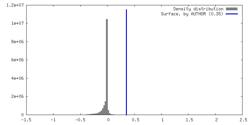
| Density |
| ||||||||||||||||||||||||||||||||||||
| Symmetry | Space group: 1 | ||||||||||||||||||||||||||||||||||||
| Details | EMDB XML:
|
 Z (Sec.)
 Y (Row.)
 X (Col.)


-Supplemental data
- Sample components
-Entire : SARS-CoV-2 spike glycoprotein in complex with neutralizing antibodies
| Entire | Name: SARS-CoV-2 spike glycoprotein in complex with neutralizing antibodies |
|---|---|
| Components |
|
-Supramolecule #1: SARS-CoV-2 spike glycoprotein in complex with neutralizing antibodies
| Supramolecule | Name: SARS-CoV-2 spike glycoprotein in complex with neutralizing antibodies type: organelle_or_cellular_component / ID: 1 / Parent: 0 / Macromolecule list: #1-#3 Details: S-Delta variant, two neutralizing antibodies RBD-chAb-15 and RBD-chAb-45 |
|---|---|
| Source (natural) | Organism: Severe acute respiratory syndrome coronavirus 2 |
| Molecular weight | Experimental: 540 KDa |
| Recombinant expression | Organism:  Homo sapiens (human) Homo sapiens (human) |
-Experimental details
-Structure determination
| Method | negative staining |
|---|---|
 Processing Processing | single particle reconstruction |
| Aggregation state | particle |
-Sample preparation
| Concentration | 0.05 mg/mL | ||||||||||||
|---|---|---|---|---|---|---|---|---|---|---|---|---|---|
| Buffer | pH: 7.6 Component:
| ||||||||||||
| Staining | Type: NEGATIVE / Material: Uranyl Formate |
- Electron microscopy
| Microscope | FEI TECNAI F20 |
|---|---|
| Image recording | Film or detector model: GATAN ULTRASCAN 4000 (4k x 4k) / Average exposure time: 1.0 sec. / Average electron dose: 30.0 e/Å2 |
| Electron beam | Acceleration voltage: 200 kV / Electron source:  FIELD EMISSION GUN FIELD EMISSION GUN |
| Electron optics | Illumination mode: FLOOD BEAM / Imaging mode: BRIGHT FIELD / Cs: 2.0 mm |
| Sample stage | Cooling holder cryogen: NITROGEN |
| Experimental equipment |  Model: Tecnai F20 / Image courtesy: FEI Company |
