 Movie
Movie Controller


+ Open data

- Basic information
| Entry |  | ||||||||||||||||||
|---|---|---|---|---|---|---|---|---|---|---|---|---|---|---|---|---|---|---|---|
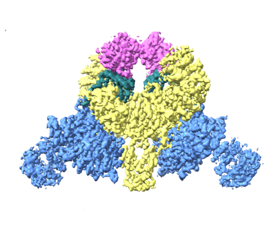
| Title | CryoEM structure of DDB1-VprBP-Vpr-UNG2(94-313) complex | ||||||||||||||||||
 Map data Map data | |||||||||||||||||||
 Sample Sample |
| ||||||||||||||||||
 Keywords Keywords | E3 ligase / HIV accessory protein / restriction factors / LIGASE / TRANSFERASE-DNA BINDING PROTEIN-VIRAL PROTEIN complex | ||||||||||||||||||
| Function / homology |  Function and homology information Function and homology informationcell competition in a multicellular organism / histone H2AT120 kinase activity / symbiont-mediated arrest of host cell cycle during G2/M transition / base-excision repair, AP site formation via deaminated base removal / uracil-DNA glycosylase / depyrimidination / Displacement of DNA glycosylase by APEX1 / single strand break repair / uracil DNA N-glycosylase activity / V(D)J recombination ...cell competition in a multicellular organism / histone H2AT120 kinase activity / symbiont-mediated arrest of host cell cycle during G2/M transition / base-excision repair, AP site formation via deaminated base removal / uracil-DNA glycosylase / depyrimidination / Displacement of DNA glycosylase by APEX1 / single strand break repair / uracil DNA N-glycosylase activity / V(D)J recombination / isotype switching / positive regulation by virus of viral protein levels in host cell / spindle assembly involved in female meiosis / epigenetic programming in the zygotic pronuclei / UV-damage excision repair / biological process involved in interaction with symbiont / regulation of mitotic cell cycle phase transition / WD40-repeat domain binding / Cul4A-RING E3 ubiquitin ligase complex / Cul4-RING E3 ubiquitin ligase complex / Cul4B-RING E3 ubiquitin ligase complex / ubiquitin ligase complex scaffold activity / negative regulation of reproductive process / negative regulation of developmental process / ribosomal small subunit binding / viral release from host cell / cullin family protein binding / ectopic germ cell programmed cell death / somatic hypermutation of immunoglobulin genes / positive regulation of viral genome replication / ubiquitin-like ligase-substrate adaptor activity / proteasomal protein catabolic process / post-translational protein modification / positive regulation of gluconeogenesis / B cell differentiation / Recognition and association of DNA glycosylase with site containing an affected pyrimidine / Cleavage of the damaged pyrimidine / nuclear estrogen receptor binding / Chromatin modifications during the maternal to zygotic transition (MZT) / symbiont-mediated activation of host apoptosis / nucleotide-excision repair / sperm end piece / regulation of circadian rhythm / protein homooligomerization / Recognition of DNA damage by PCNA-containing replication complex / base-excision repair / virion component / viral penetration into host nucleus / DNA Damage Recognition in GG-NER / fibrillar center / Dual Incision in GG-NER / Transcription-Coupled Nucleotide Excision Repair (TC-NER) / Wnt signaling pathway / Formation of TC-NER Pre-Incision Complex / Formation of Incision Complex in GG-NER / Dual incision in TC-NER / positive regulation of protein catabolic process / Gap-filling DNA repair synthesis and ligation in TC-NER / cellular response to UV / rhythmic process / host cell / Antigen processing: Ubiquitination & Proteasome degradation / site of double-strand break / sperm principal piece / Neddylation / sperm midpiece / host extracellular region / monoatomic ion transmembrane transport / ubiquitin-dependent protein catabolic process / damaged DNA binding / proteasome-mediated ubiquitin-dependent protein catabolic process / protein-macromolecule adaptor activity / chromosome, telomeric region / non-specific serine/threonine protein kinase / protein ubiquitination / protein serine kinase activity / DNA repair / apoptotic process / DNA damage response / symbiont entry into host cell / regulation of DNA-templated transcription / centrosome / negative regulation of apoptotic process / DNA-templated transcription / host cell nucleus / protein-containing complex binding / nucleolus / negative regulation of transcription by RNA polymerase II / protein-containing complex / mitochondrion / : / DNA binding / extracellular exosome / nucleoplasm / ATP binding / nucleus / cytoplasm Similarity search - Function | ||||||||||||||||||
| Biological species |  Homo sapiens (human) / Homo sapiens (human) /   Human immunodeficiency virus 1 Human immunodeficiency virus 1 | ||||||||||||||||||
| Method | single particle reconstruction / cryo EM / Resolution: 3.7 Å | ||||||||||||||||||
 Authors Authors | Wang D / Xu J | ||||||||||||||||||
| Funding support |  China, 5 items China, 5 items
| ||||||||||||||||||
 Citation Citation | Journal: To Be Published Title: Structural insights into the HIV-1 Vpr mediated ubiquitination through the Cullin-RING E3 ubiquitin ligase Authors: Wang D / Xu J / Liu Q / Xiang Y | ||||||||||||||||||
| History |
|
- Structure visualization
| Supplemental images |
|---|
- Downloads & links
-EMDB archive
| Map data | emd_31766.map.gz | 111.5 MB | EMDB map data format | |
|---|---|---|---|---|
| Header (meta data) | emd-31766-v30.xmlemd-31766.xml | 16 KB 16 KB | Display Display | EMDB header |
| Images |  emd_31766.png emd_31766.png | 118.7 KB | ||
| Filedesc metadata | emd-31766.cif.gz | 7.2 KB | ||
| Archive directory |  http://ftp.pdbj.org/pub/emdb/structures/EMD-31766ftp://ftp.pdbj.org/pub/emdb/structures/EMD-31766 http://ftp.pdbj.org/pub/emdb/structures/EMD-31766ftp://ftp.pdbj.org/pub/emdb/structures/EMD-31766 | HTTPS FTP |
-Related structure data
| Related structure data |  7v7cMC  7v7bC M: atomic model generated by this map C: citing same article ( |
|---|---|
| Similar structure data |
-Links
| EMDB pages | EMDB (EBI/PDBe) / EMDataResource |
|---|---|
| Related items in Molecule of the Month |


-Map
| File | Download / File: emd_31766.map.gz / Format: CCP4 / Size: 125 MB / Type: IMAGE STORED AS FLOATING POINT NUMBER (4 BYTES) | ||||||||||||||||||||||||||||||||||||
|---|---|---|---|---|---|---|---|---|---|---|---|---|---|---|---|---|---|---|---|---|---|---|---|---|---|---|---|---|---|---|---|---|---|---|---|---|---|
| Projections & slices | Image control
Images are generated by Spider. | ||||||||||||||||||||||||||||||||||||
| Voxel size | X=Y=Z: 1.0742 Å | ||||||||||||||||||||||||||||||||||||
| Density |
| ||||||||||||||||||||||||||||||||||||
| Symmetry | Space group: 1 | ||||||||||||||||||||||||||||||||||||
| Details | EMDB XML:
|
 Z (Sec.)
 Y (Row.)
 X (Col.)


-Supplemental data
- Sample components
-Entire : DDB1-VprBP-Vpr-UNG2(94-313) complex
| Entire | Name: DDB1-VprBP-Vpr-UNG2(94-313) complex |
|---|---|
| Components |
|
-Supramolecule #1: DDB1-VprBP-Vpr-UNG2(94-313) complex
| Supramolecule | Name: DDB1-VprBP-Vpr-UNG2(94-313) complex / type: complex / ID: 1 / Parent: 0 / Macromolecule list: all |
|---|---|
| Source (natural) | Organism: Homo sapiens (human) |
| Molecular weight | Theoretical: 0.66 kDa/nm |
-Macromolecule #1: DDB1- and CUL4-associated factor 1
| Macromolecule | Name: DDB1- and CUL4-associated factor 1 / type: protein_or_peptide / ID: 1 / Number of copies: 2 / Enantiomer: LEVO / EC number: non-specific serine/threonine protein kinase |
|---|---|
| Source (natural) | Organism: Homo sapiens (human) |
| Molecular weight | Theoretical: 169.194781 KDa |
| Recombinant expression | Organism:  Trichoplusia ni (cabbage looper) Trichoplusia ni (cabbage looper) |
| Sequence | String: MTTVVVHVDS KAELTTLLEQ WEKEHGSGQD MVPILTRMSQ LIEKETEEYR KGDPDPFDDR HPGRADPECM LGHLLRILFK NDDFMNALV NAYVMTSREP PLNTAACRLL LDIMPGLETA VVFQEKEGIV ENLFKWAREA DQPLRTYSTG LLGGAMENQD I AANYRDEN ...String: MTTVVVHVDS KAELTTLLEQ WEKEHGSGQD MVPILTRMSQ LIEKETEEYR KGDPDPFDDR HPGRADPECM LGHLLRILFK NDDFMNALV NAYVMTSREP PLNTAACRLL LDIMPGLETA VVFQEKEGIV ENLFKWAREA DQPLRTYSTG LLGGAMENQD I AANYRDEN SQLVAIVLRR LRELQLQEVA LRQENKRPSP RKLSSEPLLP LDEEAVDMDY GDMAVDVVDG DQEEASGDME IS FHLDSGH KTSSRVNSTT KPEDGGLKKN KSAKQGDREN FRKAKQKLGF SSSDPDRMFV ELSNSSWSEM SPWVIGTNYT LYP MTPAIE QRLILQYLTP LGEYQELLPI FMQLGSRELM MFYIDLKQTN DVLLTFEALK HLASLLLHNK FATEFVAHGG VQKL LEIPR PSMAATGVSM CLYYLSYNQD AMERVCMHPH NVLSDVVNYT LWLMECSHAS GCCHATMFFS ICFSFRAVLE LFDRY DGLR RLVNLISTLE ILNLEDQGAL LSDDEIFASR QTGKHTCMAL RKYFEAHLAI KLEQVKQSLQ RTEGGILVHP QPPYKA CSY THEQIVEMME FLIEYGPAQL YWEPAEVFLK LSCVQLLLQL ISIACNWKTY YARNDTVRFA LDVLAILTVV PKIQLQL AE SVDVLDEAGS TVSTVGISII LGVAEGEFFI HDAEIQKSAL QIIINCVCGP DNRISSIGKF ISGTPRRKLP QNPKSSEH T LAKMWNVVQS NNGIKVLLSL LSIKMPITDA DQIRALACKA LVGLSRSSTV RQIISKLPLF SSCQIQQLMK EPVLQDKRS DHVKFCKYAA ELIERVSGKP LLIGTDVSLA RLQKADVVAQ SRISFPEKEL LLLIRNHLIS KGLGETATVL TKEADLPMTA ASHSSAFTP VTAAASPVSL PRTPRIANGI ATRLGSHAAV GASAPSAPTA HPQPRPPQGP LALPGPSYAG NSPLIGRISF I RERPSPCN GRKIRVLRQK SDHGAYSQSP AIKKQLDRHL PSPPTLDSII TEYLREQHAR CKNPVATCPP FSLFTPHQCP EP KQRRQAP INFTSRLNRR ASFPKYGGVD GGCFDRHLIF SRFRPISVFR EANEDESGFT CCAFSARERF LMLGTCTGQL KLY NVFSGQ EEASYNCHNS AITHLEPSRD GSLLLTSATW SQPLSALWGM KSVFDMKHSF TEDHYVEFSK HSQDRVIGTK GDIA HIYDI QTGNKLLTLF NPDLANNYKR NCATFNPTDD LVLNDGVLWD VRSAQAIHKF DKFNMNISGV FHPNGLEVII NTEIW DLRT FHLLHTVPAL DQCRVVFNHT GTVMYGAMLQ ADDEDDLMEE RMKSPFGSSF RTFNATDYKP IATIDVKRNI FDLCTD TKD CYLAVIENQG SMDALNMDTV CRLYEVGRQR LAEDEDEEED QEEEEQEEED DDEDDDDTDD LDELDTDQLL EAELEED DN NENAGEDGDN DFSPSDEELA NLLEEGEDGE DEDSDADEEV ELILGDTDSS DNSDLEDDII LSLNE UniProtKB: DDB1- and CUL4-associated factor 1 |
-Macromolecule #2: DNA damage-binding protein 1
| Macromolecule | Name: DNA damage-binding protein 1 / type: protein_or_peptide / ID: 2 / Number of copies: 2 / Enantiomer: LEVO |
|---|---|
| Source (natural) | Organism: Homo sapiens (human) |
| Molecular weight | Theoretical: 127.097469 KDa |
| Recombinant expression | Organism: Trichoplusia ni (cabbage looper) |
| Sequence | String: MSYNYVVTAQ KPTAVNGCVT GHFTSAEDLN LLIAKNTRLE IYVVTAEGLR PVKEVGMYGK IAVMELFRPK GESKDLLFIL TAKYNACIL EYKQSGESID IITRAHGNVQ DRIGRPSETG IIGIIDPECR MIGLRLYDGL FKVIPLDRDN KELKAFNIRL E ELHVIDVK ...String: MSYNYVVTAQ KPTAVNGCVT GHFTSAEDLN LLIAKNTRLE IYVVTAEGLR PVKEVGMYGK IAVMELFRPK GESKDLLFIL TAKYNACIL EYKQSGESID IITRAHGNVQ DRIGRPSETG IIGIIDPECR MIGLRLYDGL FKVIPLDRDN KELKAFNIRL E ELHVIDVK FLYGCQAPTI CFVYQDPQGR HVKTYEVSLR EKEFNKGPWK QENVEAEASM VIAVPEPFGG AIIIGQESIT YH NGDKYLA IAPPIIKQST IVCHNRVDPN GSRYLLGDME GRLFMLLLEK EEQMDGTVTL KDLRVELLGE TSIAECLTYL DNG VVFVGS RLGDSQLVKL NVDSNEQGSY VVAMETFTNL GPIVDMCVVD LERQGQGQLV TCSGAFKEGS LRIIRNGIGI HEHA SIDLP GIKGLWPLRS DPNRETDDTL VLSFVGQTRV LMLNGEEVEE TELMGFVDDQ QTFFCGNVAH QQLIQITSAS VRLVS QEPK ALVSEWKEPQ AKNISVASCN SSQVVVAVGR ALYYLQIHPQ ELRQISHTEM EHEVACLDIT PLGDSNGLSP LCAIGL WTD ISARILKLPS FELLHKEMLG GEIIPRSILM TTFESSHYLL CALGDGALFY FGLNIETGLL SDRKKVTLGT QPTVLRT FR SLSTTNVFAC SDRPTVIYSS NHKLVFSNVN LKEVNYMCPL NSDGYPDSLA LANNSTLTIG TIDEIQKLHI RTVPLYES P RKICYQEVSQ CFGVLSSRIE VQDTSGGTTA LRPSASTQAL SSSVSSSKLF SSSTAPHETS FGEEVEVHNL LIIDQHTFE VLHAHQFLQN EYALSLVSCK LGKDPNTYFI VGTAMVYPEE AEPKQGRIVV FQYSDGKLQT VAEKEVKGAV YSMVEFNGKL LASINSTVR LYEWTTEKEL RTECNHYNNI MALYLKTKGD FILVGDLMRS VLLLAYKPME GNFEEIARDF NPNWMSAVEI L DDDNFLGA ENAFNLFVCQ KDSAATTDEE RQHLQEVGLF HLGEFVNVFC HGSLVMQNLG ETSTPTQGSV LFGTVNGMIG LV TSLSESW YNLLLDMQNR LNKVIKSVGK IEHSFWRSFH TERKTEPATG FIDGDLIESF LDISRPKMQE VVANLQYDDG SGM KREATA DDLIKVVEEL TRIH UniProtKB: DNA damage-binding protein 1 |
-Macromolecule #3: Protein Vpr
| Macromolecule | Name: Protein Vpr / type: protein_or_peptide / ID: 3 / Number of copies: 2 / Enantiomer: LEVO |
|---|---|
| Source (natural) | Organism: Human immunodeficiency virus 1 |
| Molecular weight | Theoretical: 11.396878 KDa |
| Recombinant expression | Organism:  |
| Sequence | String: MEQAPEDQGP QREPYNEWTL ELLEELKSEA VRHFPRIWLH NLGQHIYETY GDTWAGVEAI IRILQQLLFI HFRIGCRHSR IGVTRQRRA RNGASRS UniProtKB: Protein Vpr |
-Macromolecule #4: Uracil-DNA glycosylase
| Macromolecule | Name: Uracil-DNA glycosylase / type: protein_or_peptide / ID: 4 / Number of copies: 2 / Enantiomer: LEVO / EC number: uracil-DNA glycosylase |
|---|---|
| Source (natural) | Organism: Homo sapiens (human) |
| Molecular weight | Theoretical: 25.136654 KDa |
| Recombinant expression | Organism: |
| Sequence | String: FGESWKKHLS GEFGKPYFIK LMGFVAEERK HYTVYPPPHQ VFTWTQMCDI KDVKVVILGQ DPYHGPNQAH GLCFSVQRPV PPPPSLENI YKELSTDIED FVHPGHGDLS GWAKQGVLLL NAVLTVRAHQ ANSHKERGWE QFTDAVVSWL NQNSNGLVFL L WGSYAQKK ...String: FGESWKKHLS GEFGKPYFIK LMGFVAEERK HYTVYPPPHQ VFTWTQMCDI KDVKVVILGQ DPYHGPNQAH GLCFSVQRPV PPPPSLENI YKELSTDIED FVHPGHGDLS GWAKQGVLLL NAVLTVRAHQ ANSHKERGWE QFTDAVVSWL NQNSNGLVFL L WGSYAQKK GSAIDRKRHH VLQTAHPSPL SVYRGFFGCR HFSKTNELLQ KSGKKPIDWK EL UniProtKB: Uracil-DNA glycosylase |
-Experimental details
-Structure determination
| Method | cryo EM |
|---|---|
 Processing Processing | single particle reconstruction |
| Aggregation state | particle |
-Sample preparation
| Buffer | pH: 7.5 |
|---|---|
| Vitrification | Cryogen name: ETHANE |
- Electron microscopy
| Microscope | FEI TITAN KRIOS |
|---|---|
| Image recording | Film or detector model: GATAN K3 (6k x 4k) / Average electron dose: 50.0 e/Å2 |
| Electron beam | Acceleration voltage: 300 kV / Electron source:  FIELD EMISSION GUN FIELD EMISSION GUN |
| Electron optics | Illumination mode: FLOOD BEAM / Imaging mode: BRIGHT FIELD |
| Experimental equipment |  Model: Titan Krios / Image courtesy: FEI Company |
-Image processing
| Startup model | Type of model: OTHER |
|---|---|
| Final reconstruction | Resolution.type: BY AUTHOR / Resolution: 3.7 Å / Resolution method: FSC 0.143 CUT-OFF / Software - Name: RELION / Number images used: 145568 |
| Initial angle assignment | Type: MAXIMUM LIKELIHOOD / Software - Name: RELION |
| Final angle assignment | Type: MAXIMUM LIKELIHOOD / Software - Name: RELION |
-Atomic model buiding 1
| Refinement | Protocol: RIGID BODY FIT |
|---|---|
| Output model | PDB-7v7c: |
