 ムービー
ムービー コントローラー


+ データを開く

- 基本情報
基本情報
| 登録情報 | データベース: EMDB / ID: EMD-3128 | |||||||||
|---|---|---|---|---|---|---|---|---|---|---|
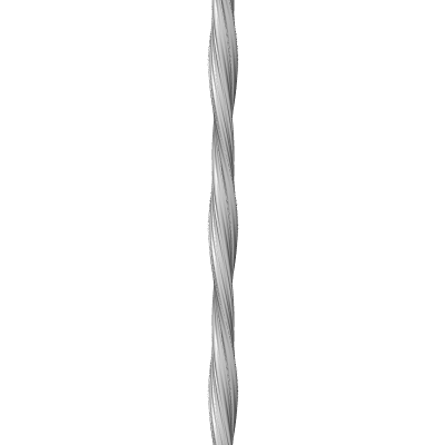
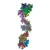
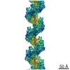
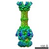
| タイトル | Structure of a cross-beta amyloid fibril from IGSNVVTWYQQL peptide of AL-DIA immunoglobulin light chain by cryo-EM and fiber diffraction | |||||||||
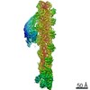
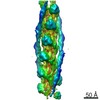
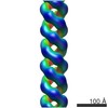
 マップデータ マップデータ | Reconstruction morphology 1 of a left-handed amyloid-like fibril of the IGSNVVTWYQQL fragment of an immunoglobulin light chain | |||||||||
 試料 試料 |
| |||||||||
 キーワード キーワード | AL amyloidosis / cryo electron microscopy / steric zipper / three dimensional reconstruction | |||||||||
| 生物種 |  Homo sapiens (ヒト) Homo sapiens (ヒト) | |||||||||
| 手法 | らせん対称体再構成法 / クライオ電子顕微鏡法 / 解像度: 8.3 Å | |||||||||
 データ登録者 データ登録者 | Schmidt A / Annamalai K / Schmidt M / Grigorieff N / Fandrich M | |||||||||
 引用 引用 | ジャーナル: Proc Natl Acad Sci U S A / 年: 2016 タイトル: Cryo-EM reveals the steric zipper structure of a light chain-derived amyloid fibril. 著者: Andreas Schmidt / Karthikeyan Annamalai / Matthias Schmidt / Nikolaus Grigorieff / Marcus Fändrich /   要旨: Amyloid fibrils are proteinaceous aggregates associated with diseases in humans and animals. The fibrils are defined by intermolecular interactions between the fibril-forming polypeptide chains, but ...Amyloid fibrils are proteinaceous aggregates associated with diseases in humans and animals. The fibrils are defined by intermolecular interactions between the fibril-forming polypeptide chains, but it has so far remained difficult to reveal the assembly of the peptide subunits in a full-scale fibril. Using electron cryomicroscopy (cryo-EM), we present a reconstruction of a fibril formed from the pathogenic core of an amyloidogenic immunoglobulin (Ig) light chain. The fibril density shows a lattice-like assembly of face-to-face packed peptide dimers that corresponds to the structure of steric zippers in peptide crystals. Interpretation of the density map with a molecular model enabled us to identify the intermolecular interactions between the peptides and rationalize the hierarchical structure of the fibril based on simple chemical principles. | |||||||||
| 履歴 |
|
- 構造の表示
構造の表示
| ムービー |
 ムービービューア ムービービューア |
|---|---|
| 構造ビューア | EMマップ: SurfViewMolmilJmol/JSmol |
| 添付画像 |
- ダウンロードとリンク
-EMDBアーカイブ
| マップデータ | emd_3128.map.gz | 2.7 MB | EMDBマップデータ形式 | |
|---|---|---|---|---|
| ヘッダ (付随情報) | emd-3128-v30.xmlemd-3128.xml | 9.8 KB 9.8 KB | 表示 表示 | EMDBヘッダ |
| FSC (解像度算出) | emd_3128_fsc.xml | 11.8 KB | 表示 | FSCデータファイル |
| 画像 |  emd_3128.png emd_3128.png | 16.8 KB | ||
| アーカイブディレクトリ |  http://ftp.pdbj.org/pub/emdb/structures/EMD-3128ftp://ftp.pdbj.org/pub/emdb/structures/EMD-3128 http://ftp.pdbj.org/pub/emdb/structures/EMD-3128ftp://ftp.pdbj.org/pub/emdb/structures/EMD-3128 | HTTPS FTP |
-検証レポート
| 文書・要旨 | emd_3128_validation.pdf.gz | 219.4 KB | 表示 | EMDB検証レポート |
|---|---|---|---|---|
| 文書・詳細版 | emd_3128_full_validation.pdf.gz | 218.4 KB | 表示 | |
| XML形式データ | emd_3128_validation.xml.gz | 10.5 KB | 表示 | |
| アーカイブディレクトリ | https://ftp.pdbj.org/pub/emdb/validation_reports/EMD-3128ftp://ftp.pdbj.org/pub/emdb/validation_reports/EMD-3128 | HTTPS FTP |
-関連構造データ

-リンク
| EMDBのページ | EMDB (EBI/PDBe) / EMDataResource |
|---|---|
| 「今月の分子」の関連する項目 |

-マップ
| ファイル | ダウンロード / ファイル: emd_3128.map.gz / 形式: CCP4 / 大きさ: 2.9 MB / タイプ: IMAGE STORED AS FLOATING POINT NUMBER (4 BYTES) | ||||||||||||||||||||||||||||||||||||||||||||||||||||||||||||
|---|---|---|---|---|---|---|---|---|---|---|---|---|---|---|---|---|---|---|---|---|---|---|---|---|---|---|---|---|---|---|---|---|---|---|---|---|---|---|---|---|---|---|---|---|---|---|---|---|---|---|---|---|---|---|---|---|---|---|---|---|---|
| 注釈 | Reconstruction morphology 1 of a left-handed amyloid-like fibril of the IGSNVVTWYQQL fragment of an immunoglobulin light chain | ||||||||||||||||||||||||||||||||||||||||||||||||||||||||||||
| 投影像・断面図 | 画像のコントロール
画像は Spider により作成 これらの図は立方格子座標系で作成されたものです | ||||||||||||||||||||||||||||||||||||||||||||||||||||||||||||
| ボクセルのサイズ | X=Y=Z: 2.11 Å | ||||||||||||||||||||||||||||||||||||||||||||||||||||||||||||
| 密度 |
| ||||||||||||||||||||||||||||||||||||||||||||||||||||||||||||
| 対称性 | 空間群: 1 | ||||||||||||||||||||||||||||||||||||||||||||||||||||||||||||
| 詳細 | EMDB XML:
CCP4マップ ヘッダ情報:
| ||||||||||||||||||||||||||||||||||||||||||||||||||||||||||||
 Z (Sec.)
 Y (Row.)
 X (Col.)


-添付データ
- 試料の構成要素
-全体 : Amyloidogenic Fragment IGSNVVTWYQQL of Immunoglobulin Light Chain...
| 全体 | 名称: Amyloidogenic Fragment IGSNVVTWYQQL of Immunoglobulin Light Chain of Human AL Patient |
|---|---|
| 要素 |
|
-超分子 #1000: Amyloidogenic Fragment IGSNVVTWYQQL of Immunoglobulin Light Chain...
| 超分子 | 名称: Amyloidogenic Fragment IGSNVVTWYQQL of Immunoglobulin Light Chain of Human AL Patient タイプ: sample / ID: 1000 / 詳細: The sample forms long amyloid-like fibrils. / Number unique components: 1 |
|---|
-分子 #1: Immunoglobulin Light Chain
| 分子 | 名称: Immunoglobulin Light Chain / タイプ: protein_or_peptide / ID: 1 / 組換発現: No / データベース: NCBI |
|---|---|
| 由来(天然) | 生物種: Homo sapiens (ヒト) / 別称: Human / 組織: Blood / 細胞: Plasma Cell / 細胞中の位置: extracellular |
-実験情報
-構造解析
| 手法 | クライオ電子顕微鏡法 |
|---|---|
 解析 解析 | らせん対称体再構成法 |
| 試料の集合状態 | filament |
-試料調製
| 濃度 | 5.0 mg/mL |
|---|---|
| 緩衝液 | pH: 8 / 詳細: 50mM Tris-HCL |
| グリッド | 詳細: C-flat 1.2/1.3-2C 400 mesh grids, glow discharged |
| 凍結 | 凍結剤: ETHANE / チャンバー内湿度: 50 % / チャンバー内温度: 100 K / 装置: GATAN CRYOPLUNGE 3 手法: Incubation of 0.014 mg/ml fibril solution on glow discharged holey carbon grid for 30 seconds and backside blotting for 4 seconds before plunging. |
- 電子顕微鏡法
| 顕微鏡 | FEI TECNAI F20 |
|---|---|
| 温度 | 平均: 100 K |
| 日付 | 2012年11月12日 |
| 撮影 | カテゴリ: CCD フィルム・検出器のモデル: FEI FALCON I (4k x 4k) 実像数: 10 / 平均電子線量: 25 e/Å2 / ビット/ピクセル: 16 |
| 電子線 | 加速電圧: 200 kV / 電子線源:  FIELD EMISSION GUN FIELD EMISSION GUN |
| 電子光学系 | 倍率(補正後): 66350.7 / 照射モード: FLOOD BEAM / 撮影モード: BRIGHT FIELD / Cs: 2.0 mm / 最大 デフォーカス(公称値): 5.0 µm / 最小 デフォーカス(公称値): 1.0 µm / 倍率(公称値): 50000 |
| 試料ステージ | 試料ホルダーモデル: GATAN LIQUID NITROGEN |
| 実験機器 |  モデル: Tecnai F20 / 画像提供: FEI Company |
-画像解析
| 最終 再構成 | 想定した対称性 - らせんパラメータ - Δz: 4.69 Å 想定した対称性 - らせんパラメータ - ΔΦ: 1.464 ° 想定した対称性 - らせんパラメータ - 軸対称性: C2 (2回回転対称) アルゴリズム: OTHER / 解像度のタイプ: BY AUTHOR / 解像度: 8.3 Å / 解像度の算出法: OTHER / ソフトウェア - 名称: Frealix 詳細: Final map was calculated from eleven single fibril reconstructions. |
|---|---|
| CTF補正 | 詳細: Defocus estimated for each helical subunit |
| FSC曲線 (解像度の算出) | 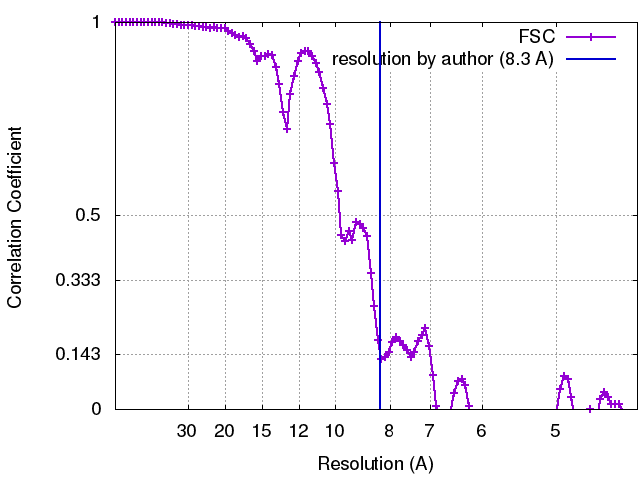 |
