 Movie
Movie Controller


+ Open data

- Basic information
| Entry | Database: EMDB / ID: EMD-3124 | |||||||||
|---|---|---|---|---|---|---|---|---|---|---|
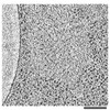
| Title | Cryo-tomogram of an isolated fibrillar [PSI+] prion assembly | |||||||||
 Map data Map data | Reconstruction of an isolated amyloid assembly | |||||||||
 Sample Sample |
| |||||||||
| Biological species |  | |||||||||
| Method | electron tomography / cryo EM | |||||||||
 Authors Authors | O'Driscoll J / Clare D / Saibil H | |||||||||
 Citation Citation | Journal: J Cell Biol / Year: 2015 Title: Prion aggregate structure in yeast cells is determined by the Hsp104-Hsp110 disaggregase machinery. Authors: Jonathan O'Driscoll / Daniel Clare / Helen Saibil /  Abstract: Prions consist of misfolded proteins that have adopted an infectious amyloid conformation. In vivo, prion biogenesis is intimately associated with the protein quality control machinery. Using ...Prions consist of misfolded proteins that have adopted an infectious amyloid conformation. In vivo, prion biogenesis is intimately associated with the protein quality control machinery. Using electron tomography, we probed the effects of the heat shock protein Hsp70 chaperone system on the structure of a model yeast [PSI+] prion in situ. Individual Hsp70 deletions shift the balance between fibril assembly and disassembly, resulting in a variable shell of nonfibrillar, but still immobile, aggregates at the surface of the [PSI+] prion deposits. Both Hsp104 (an Hsp100 disaggregase) and Sse1 (the major yeast form of Hsp110) were localized to this surface shell of [PSI+] deposits in the deletion mutants. Elevation of Hsp104 expression promoted the appearance of this novel, nonfibrillar form of the prion aggregate. Moreover, Sse1 was found to regulate prion fibril length. Our studies reveal a key role for Sse1 (Hsp110), in cooperation with Hsp104, in regulating the length and assembly state of [PSI+] prion fibrils in vivo. | |||||||||
| History |
|
- Structure visualization
| Movie |
 Movie viewer Movie viewer |
|---|---|
| Structure viewer | EM map: SurfViewMolmilJmol/JSmol |
| Supplemental images |

- Downloads & links
-EMDB archive
| Map data | emd_3124.map.gz | 183.8 MB | EMDB map data format | |
|---|---|---|---|---|
| Header (meta data) | emd-3124-v30.xmlemd-3124.xml | 8.6 KB 8.6 KB | Display Display | EMDB header |
| Images | emd_3124.tif | 244.3 KB | ||
| Archive directory |  http://ftp.pdbj.org/pub/emdb/structures/EMD-3124ftp://ftp.pdbj.org/pub/emdb/structures/EMD-3124 http://ftp.pdbj.org/pub/emdb/structures/EMD-3124ftp://ftp.pdbj.org/pub/emdb/structures/EMD-3124 | HTTPS FTP |
-Related structure data

-Links
| EMDB pages | EMDB (EBI/PDBe) / EMDataResource |
|---|
-Map
| File | Download / File: emd_3124.map.gz / Format: CCP4 / Size: 424.9 MB / Type: IMAGE STORED AS SIGNED BYTE | ||||||||||||||||||||||||||||||||||||||||||||||||||||||||||||
|---|---|---|---|---|---|---|---|---|---|---|---|---|---|---|---|---|---|---|---|---|---|---|---|---|---|---|---|---|---|---|---|---|---|---|---|---|---|---|---|---|---|---|---|---|---|---|---|---|---|---|---|---|---|---|---|---|---|---|---|---|---|
| Annotation | Reconstruction of an isolated amyloid assembly | ||||||||||||||||||||||||||||||||||||||||||||||||||||||||||||
| Voxel size | X=Y=Z: 11.41 Å | ||||||||||||||||||||||||||||||||||||||||||||||||||||||||||||
| Density |
| ||||||||||||||||||||||||||||||||||||||||||||||||||||||||||||
| Symmetry | Space group: 1 | ||||||||||||||||||||||||||||||||||||||||||||||||||||||||||||
| Details | EMDB XML:
CCP4 map header:
| ||||||||||||||||||||||||||||||||||||||||||||||||||||||||||||
-Supplemental data
- Sample components
-Entire : Isolated yeast prion fibril assembly
| Entire | Name: Isolated yeast prion fibril assembly |
|---|---|
| Components |
|
-Supramolecule #1000: Isolated yeast prion fibril assembly
| Supramolecule | Name: Isolated yeast prion fibril assembly / type: sample / ID: 1000 / Number unique components: 1 |
|---|
-Supramolecule #1: Yeast Prion aggregate deposit
| Supramolecule | Name: Yeast Prion aggregate deposit / type: organelle_or_cellular_component / ID: 1 / Name.synonym: [PSI+] / Recombinant expression: No / Database: NCBI |
|---|---|
| Source (natural) | Organism: |
-Experimental details
-Structure determination
| Method | cryo EM |
|---|---|
 Processing Processing | electron tomography |
| Aggregation state | cell |
-Sample preparation
| Buffer | pH: 7.4 / Details: 50 mM Tris |
|---|---|
| Grid | Details: 200 mesh c-flat carbon support film augmented with an additional 5 nm thin continuous carbon film |
| Vitrification | Cryogen name: ETHANE / Chamber temperature: 78 K / Instrument: HOMEMADE PLUNGER / Method: Blot for 8 seconds before plunging |
- Electron microscopy
| Microscope | FEI POLARA 300 |
|---|---|
| Temperature | Min: 76 K / Max: 78 K / Average: 77 K |
| Date | Jun 21, 2013 |
| Image recording | Category: CCD / Film or detector model: GATAN MULTISCAN / Number real images: 81 / Average electron dose: 1 e/Å2 Details: Each image was binned by 2 from the original image stack collected on the CCD camera Bits/pixel: 8 |
| Electron beam | Acceleration voltage: 300 kV / Electron source:  FIELD EMISSION GUN FIELD EMISSION GUN |
| Electron optics | Illumination mode: FLOOD BEAM / Imaging mode: BRIGHT FIELD / Cs: 2.3 mm / Nominal defocus max: 10.0 µm / Nominal defocus min: 6.0 µm / Nominal magnification: 20000 |
| Sample stage | Specimen holder: Liquid nitrogen cooled / Specimen holder model: OTHER / Tilt series - Axis1 - Min angle: -60 ° / Tilt series - Axis1 - Max angle: 60 ° / Tilt series - Axis1 - Angle increment: 1.5 ° |
| Experimental equipment |  Model: Tecnai Polara / Image courtesy: FEI Company |
-Image processing
| Details | Reconstructed using IMOD, no CTF correction was performed. The final reconstruction was binned by 2. The reconstruction was NAD filtered. |
|---|---|
| Final reconstruction | Software - Name: IMOD / Number images used: 75 |
