 Movie
Movie Controller


+ Open data

- Basic information
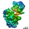
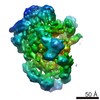
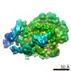
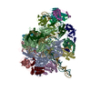
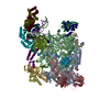
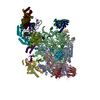
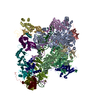
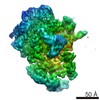
| Entry | Database: EMDB / ID: EMD-3115 | |||||||||
|---|---|---|---|---|---|---|---|---|---|---|
| Title | the P-lobe of RNA polymerase II pre-initiation complex | |||||||||
 Map data Map data | refined P-lobe of RNA polymerase II pre-initiation complex | |||||||||
 Sample Sample |
| |||||||||
 Keywords Keywords | transcription / pre-initiation complex / RNA polymerase / TFIIE / TFIIH / TFIIB / TBP / TFIIF | |||||||||
| Biological species |  | |||||||||
| Method | single particle reconstruction / cryo EM / Resolution: 6.0 Å | |||||||||
 Authors Authors | Murakami K / Tsai K-L / Kalisman N / Bushnell DA / Asturias FJ / Kornberg RD | |||||||||
 Citation Citation | Journal: Proc Natl Acad Sci U S A / Year: 2015 Title: Structure of an RNA polymerase II preinitiation complex. Authors: Kenji Murakami / Kuang-Lei Tsai / Nir Kalisman / David A Bushnell / Francisco J Asturias / Roger D Kornberg /   Abstract: The structure of a 33-protein, 1.5-MDa RNA polymerase II preinitiation complex (PIC) was determined by cryo-EM and image processing at a resolution of 6-11 Å. Atomic structures of over 50% of the ...The structure of a 33-protein, 1.5-MDa RNA polymerase II preinitiation complex (PIC) was determined by cryo-EM and image processing at a resolution of 6-11 Å. Atomic structures of over 50% of the mass were fitted into the electron density map in a manner consistent with protein-protein cross-links previously identified by mass spectrometry. The resulting model of the PIC confirmed the main conclusions from previous cryo-EM at lower resolution, including the association of promoter DNA only with general transcription factors and not with the polymerase. Electron density due to DNA was identifiable by the grooves of the double helix and exhibited sharp bends at points downstream of the TATA box, with an important consequence: The DNA at the downstream end coincides with the DNA in a transcribing polymerase. The structure of the PIC is therefore conducive to promoter melting, start-site scanning, and the initiation of transcription. | |||||||||
| History |
|
- Structure visualization
| Movie |
 Movie viewer Movie viewer |
|---|---|
| Structure viewer | EM map: SurfViewMolmilJmol/JSmol |
| Supplemental images |

- Downloads & links
-EMDB archive
| Map data | emd_3115.map.gz | 95.9 MB | EMDB map data format | |
|---|---|---|---|---|
| Header (meta data) | emd-3115-v30.xmlemd-3115.xml | 8.3 KB 8.3 KB | Display Display | EMDB header |
| Images |  emd_3115.png emd_3115.png | 245.8 KB | ||
| Archive directory |  http://ftp.pdbj.org/pub/emdb/structures/EMD-3115ftp://ftp.pdbj.org/pub/emdb/structures/EMD-3115 http://ftp.pdbj.org/pub/emdb/structures/EMD-3115ftp://ftp.pdbj.org/pub/emdb/structures/EMD-3115 | HTTPS FTP |
-Related structure data


-Links
| EMDB pages | EMDB (EBI/PDBe) / EMDataResource |
|---|
-Map
| File | Download / File: emd_3115.map.gz / Format: CCP4 / Size: 104.7 MB / Type: IMAGE STORED AS FLOATING POINT NUMBER (4 BYTES) | ||||||||||||||||||||||||||||||||||||||||||||||||||||||||||||
|---|---|---|---|---|---|---|---|---|---|---|---|---|---|---|---|---|---|---|---|---|---|---|---|---|---|---|---|---|---|---|---|---|---|---|---|---|---|---|---|---|---|---|---|---|---|---|---|---|---|---|---|---|---|---|---|---|---|---|---|---|---|
| Annotation | refined P-lobe of RNA polymerase II pre-initiation complex | ||||||||||||||||||||||||||||||||||||||||||||||||||||||||||||
| Projections & slices | Image control
Images are generated by Spider. | ||||||||||||||||||||||||||||||||||||||||||||||||||||||||||||
| Voxel size | X=Y=Z: 1.315 Å | ||||||||||||||||||||||||||||||||||||||||||||||||||||||||||||
| Density |
| ||||||||||||||||||||||||||||||||||||||||||||||||||||||||||||
| Symmetry | Space group: 1 | ||||||||||||||||||||||||||||||||||||||||||||||||||||||||||||
| Details | EMDB XML:
CCP4 map header:
| ||||||||||||||||||||||||||||||||||||||||||||||||||||||||||||
 Z (Sec.)
 Y (Row.)
 X (Col.)


-Supplemental data
- Sample components
-Entire : refined P-lobe of yeast RNA polymerase II pre-initiation complex
| Entire | Name: refined P-lobe of yeast RNA polymerase II pre-initiation complex |
|---|---|
| Components |
|
-Supramolecule #1000: refined P-lobe of yeast RNA polymerase II pre-initiation complex
| Supramolecule | Name: refined P-lobe of yeast RNA polymerase II pre-initiation complex type: sample / ID: 1000 / Number unique components: 1 |
|---|---|
| Molecular weight | Experimental: 1.5 MDa |
-Macromolecule #1: RNA polymerase II pre-initiation complex
| Macromolecule | Name: RNA polymerase II pre-initiation complex / type: protein_or_peptide / ID: 1 / Name.synonym: PIC / Recombinant expression: No |
|---|---|
| Source (natural) | Organism: |
| Molecular weight | Experimental: 1.5 MDa |
-Experimental details
-Structure determination
| Method | cryo EM |
|---|---|
 Processing Processing | single particle reconstruction |
| Aggregation state | particle |
-Sample preparation
| Concentration | 0.3 mg/mL |
|---|---|
| Buffer | pH: 7.6 Details: 20 mM HEPES (pH7.6), 5 mM DTT, 2 mM Mg(OAc)2, and 40 mM KOAc |
| Grid | Details: 3uL was transferred to Quantifoil and flash frozen in liquid ethane with a Vitrobot. |
| Vitrification | Cryogen name: ETHANE / Chamber humidity: 100 % / Chamber temperature: 120 K / Instrument: FEI VITROBOT MARK III |
- Electron microscopy
| Microscope | FEI TITAN KRIOS |
|---|---|
| Date | Nov 14, 2014 |
| Image recording | Category: CCD / Film or detector model: GATAN K2 SUMMIT (4k x 4k) / Number real images: 2564 / Average electron dose: 40 e/Å2 |
| Tilt angle min | 0 |
| Tilt angle max | 0 |
| Electron beam | Acceleration voltage: 300 kV / Electron source:  FIELD EMISSION GUN FIELD EMISSION GUN |
| Electron optics | Illumination mode: FLOOD BEAM / Imaging mode: BRIGHT FIELD / Cs: 2.70 mm / Nominal defocus max: 3.0 µm / Nominal defocus min: 1.0 µm / Nominal magnification: 22500 |
| Sample stage | Specimen holder model: FEI TITAN KRIOS AUTOGRID HOLDER |
| Experimental equipment |  Model: Titan Krios / Image courtesy: FEI Company |
-Image processing
| CTF correction | Details: Sparx |
|---|---|
| Final reconstruction | Applied symmetry - Point group: C1 (asymmetric) / Algorithm: OTHER / Resolution.type: BY AUTHOR / Resolution: 6.0 Å / Resolution method: OTHER / Software - Name: Sparx / Number images used: 37328 |
