 Movie
Movie Controller


+ Open data

- Basic information
| Entry | Database: EMDB / ID: EMD-30832 | |||||||||
|---|---|---|---|---|---|---|---|---|---|---|
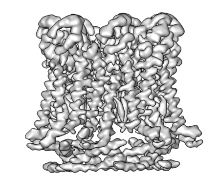
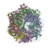
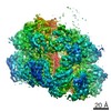
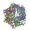
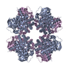
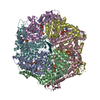
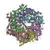
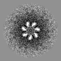
| Title | CALHM1 close state with ordered CTH | |||||||||
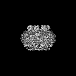
 Map data Map data | EM map | |||||||||
 Sample Sample |
| |||||||||
 Keywords Keywords | close state / MEMBRANE PROTEIN | |||||||||
| Function / homology | Calcium homeostasis modulator family / Calcium homeostasis modulator / ATP export / voltage-gated calcium channel activity / plasma membrane / Calcium homeostasis modulator 1 Function and homology information Function and homology information | |||||||||
| Biological species |  | |||||||||
| Method | single particle reconstruction / cryo EM / Resolution: 3.2 Å | |||||||||
 Authors Authors | Ren Y / Yang X | |||||||||
| Funding support |  China, 1 items China, 1 items
| |||||||||
 Citation Citation | Journal: J Biol Chem / Year: 2022 Title: Cryo-EM structure of the heptameric calcium homeostasis modulator 1 channel. Authors: Yue Ren / Yang Li / Yaojie Wang / Tianlei Wen / Xuhang Lu / Shenghai Chang / Xing Zhang / Yuequan Shen / Xue Yang / Abstract: Calcium homeostasis modulator 1 (CALHM1) is a voltage- and Ca-gated ATP channel that plays an important role in neuronal signaling. However, as the previously reported CALHM structures are all in the ...Calcium homeostasis modulator 1 (CALHM1) is a voltage- and Ca-gated ATP channel that plays an important role in neuronal signaling. However, as the previously reported CALHM structures are all in the ATP-conducting state, the gating mechanism of ATP permeation is still elusive. Here, we report cryo-EM reconstructions of two Danio rerio CALHM1 heptamers with ordered or flexible long C-terminal helices at resolutions of 3.2 Å and 2.9 Å, respectively, and one D. rerio CALHM1 octamer with flexible long C-terminal helices at a resolution of 3.5 Å. Structural analysis shows that the heptameric CALHM1s are in an ATP-nonconducting state with a central pore diameter of approximately 6.6 Å. Compared with those inside the octameric CALHM1, the N-helix inside the heptameric CALHM1 is in the "down" position to avoid steric clashing with the adjacent TM1 helix. Molecular dynamics simulations show that as the N-helix moves from the "down" position to the "up" position, the pore size of ATP molecule permeation increases significantly. Our results provide important information for elucidating the mechanism of ATP molecule permeation in the CALHM1 channel. | |||||||||
| History |
|
- Structure visualization
| Movie |
Movie viewer |
|---|---|
| Structure viewer | EM map: SurfViewMolmilJmol/JSmol |
| Supplemental images |
- Downloads & links
-EMDB archive
| Map data | emd_30832.map.gz | 33.5 MB | EMDB map data format | |
|---|---|---|---|---|
| Header (meta data) | emd-30832-v30.xmlemd-30832.xml | 10.5 KB 10.5 KB | Display Display | EMDB header |
| Images |  emd_30832.png emd_30832.png | 96.1 KB | ||
| Filedesc metadata | emd-30832.cif.gz | 5.4 KB | ||
| Archive directory |  http://ftp.pdbj.org/pub/emdb/structures/EMD-30832ftp://ftp.pdbj.org/pub/emdb/structures/EMD-30832 http://ftp.pdbj.org/pub/emdb/structures/EMD-30832ftp://ftp.pdbj.org/pub/emdb/structures/EMD-30832 | HTTPS FTP |
-Validation report
| Summary document | emd_30832_validation.pdf.gz | 498.2 KB | Display | EMDB validaton report |
|---|---|---|---|---|
| Full document | emd_30832_full_validation.pdf.gz | 497.7 KB | Display | |
| Data in XML | emd_30832_validation.xml.gz | 6.4 KB | Display | |
| Data in CIF | emd_30832_validation.cif.gz | 7.4 KB | Display | |
| Arichive directory | https://ftp.pdbj.org/pub/emdb/validation_reports/EMD-30832ftp://ftp.pdbj.org/pub/emdb/validation_reports/EMD-30832 | HTTPS FTP |
-Related structure data
| Related structure data |  7dseMC  7dscC  7dsdC M: atomic model generated by this map C: citing same article ( |
|---|---|
| Similar structure data |
-Links
| EMDB pages | EMDB (EBI/PDBe) / EMDataResource |
|---|
-Map
| File | Download / File: emd_30832.map.gz / Format: CCP4 / Size: 67 MB / Type: IMAGE STORED AS FLOATING POINT NUMBER (4 BYTES) | ||||||||||||||||||||||||||||||||||||||||||||||||||||||||||||||||||||
|---|---|---|---|---|---|---|---|---|---|---|---|---|---|---|---|---|---|---|---|---|---|---|---|---|---|---|---|---|---|---|---|---|---|---|---|---|---|---|---|---|---|---|---|---|---|---|---|---|---|---|---|---|---|---|---|---|---|---|---|---|---|---|---|---|---|---|---|---|---|
| Annotation | EM map | ||||||||||||||||||||||||||||||||||||||||||||||||||||||||||||||||||||
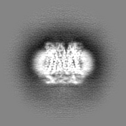
| Projections & slices | Image control
Images are generated by Spider. | ||||||||||||||||||||||||||||||||||||||||||||||||||||||||||||||||||||
| Voxel size | X=Y=Z: 1.014 Å | ||||||||||||||||||||||||||||||||||||||||||||||||||||||||||||||||||||
| Density |
| ||||||||||||||||||||||||||||||||||||||||||||||||||||||||||||||||||||
| Symmetry | Space group: 1 | ||||||||||||||||||||||||||||||||||||||||||||||||||||||||||||||||||||
| Details | EMDB XML:
CCP4 map header:
| ||||||||||||||||||||||||||||||||||||||||||||||||||||||||||||||||||||
 Z (Sec.)
 Y (Row.)
 X (Col.)


-Supplemental data
- Sample components
-Entire : CALHM1
| Entire | Name: CALHM1 |
|---|---|
| Components |
|
-Supramolecule #1: CALHM1
| Supramolecule | Name: CALHM1 / type: organelle_or_cellular_component / ID: 1 / Parent: 0 / Macromolecule list: #1 |
|---|---|
| Source (natural) | Organism: |
-Macromolecule #1: Calcium homeostasis modulator 1
| Macromolecule | Name: Calcium homeostasis modulator 1 / type: protein_or_peptide / ID: 1 / Number of copies: 7 / Enantiomer: LEVO |
|---|---|
| Source (natural) | Organism: |
| Molecular weight | Theoretical: 41.213645 KDa |
| Recombinant expression | Organism:  Homo sapiens (human) Homo sapiens (human) |
| Sequence | String: LEQKLISEED LRSDKFRIMV QFLQANQESF MNGICGIMAL ASAQMYSSFE FTCPCLPDYN YAYGIGILIV PPIWFFLLGY VMNNNISVL TEEWKRPVGK RSKDPAVLRY MFSSMTQRAL IAPAVWIAVT LMDGKSFLCA FSPTADLSEF VNESYQSLSQ K ELLKIQAK ...String: LEQKLISEED LRSDKFRIMV QFLQANQESF MNGICGIMAL ASAQMYSSFE FTCPCLPDYN YAYGIGILIV PPIWFFLLGY VMNNNISVL TEEWKRPVGK RSKDPAVLRY MFSSMTQRAL IAPAVWIAVT LMDGKSFLCA FSPTADLSEF VNESYQSLSQ K ELLKIQAK IPCKDIFEEH EIISREAATR YIRCLSQACG WTFLMVITLV AFLVRAIRPC FTQAAFLKTK YWSHYIDTER KL FDETCKE HAKSFAKVCI QQYFESISGE IVSQLPQSPA KKGKGNKDED GEKQKSDEER LLGIRKEGDM NKVLWNWHTC KPP LLLSKR TEEMNGHAHL DTHSLTDERH TKKKAVVYYS KV UniProtKB: Calcium homeostasis modulator 1 |
-Macromolecule #2: 2-acetamido-2-deoxy-beta-D-glucopyranose
| Macromolecule | Name: 2-acetamido-2-deoxy-beta-D-glucopyranose / type: ligand / ID: 2 / Number of copies: 7 / Formula: NAG |
|---|---|
| Molecular weight | Theoretical: 221.208 Da |
| Chemical component information |  ChemComp-NAG: |
-Experimental details
-Structure determination
| Method | cryo EM |
|---|---|
 Processing Processing | single particle reconstruction |
| Aggregation state | 2D array |
-Sample preparation
| Concentration | 10 mg/mL |
|---|---|
| Buffer | pH: 7.5 |
| Grid | Model: Quantifoil / Support film - Material: CARBON / Support film - topology: CONTINUOUS |
| Vitrification | Cryogen name: ETHANE / Chamber humidity: 100 % / Chamber temperature: 281 K / Instrument: FEI VITROBOT MARK IV |
- Electron microscopy
| Microscope | FEI TITAN KRIOS |
|---|---|
| Image recording | Film or detector model: GATAN K2 SUMMIT (4k x 4k) / Detector mode: COUNTING / Average electron dose: 50.0 e/Å2 |
| Electron beam | Acceleration voltage: 300 kV / Electron source:  FIELD EMISSION GUN FIELD EMISSION GUN |
| Electron optics | Illumination mode: FLOOD BEAM / Imaging mode: BRIGHT FIELD / Nominal defocus max: 2.5 µm / Nominal defocus min: 1.5 µm |
| Sample stage | Cooling holder cryogen: NITROGEN |
| Experimental equipment |  Model: Titan Krios / Image courtesy: FEI Company |
-Image processing
| Startup model | Type of model: OTHER / Details: Ab into from cryosparc v2 |
|---|---|
| Final reconstruction | Resolution.type: BY AUTHOR / Resolution: 3.2 Å / Resolution method: FSC 0.143 CUT-OFF / Number images used: 12671 |
| Initial angle assignment | Type: ANGULAR RECONSTITUTION |
| Final angle assignment | Type: NOT APPLICABLE |
