mRNA 5'-UTR binding / large ribosomal subunit / transferase activity / 5S rRNA binding / ribosomal large subunit assembly / small ribosomal subunit / small ribosomal subunit rRNA binding / cytosolic small ribosomal subunit / large ribosomal subunit rRNA binding / cytosolic large ribosomal subunit ...mRNA 5'-UTR binding / large ribosomal subunit / transferase activity / 5S rRNA binding / ribosomal large subunit assembly / small ribosomal subunit / small ribosomal subunit rRNA binding / cytosolic small ribosomal subunit / large ribosomal subunit rRNA binding / cytosolic large ribosomal subunit / cytoplasmic translation / tRNA binding / rRNA binding / structural constituent of ribosome / ribosome / translation / ribonucleoprotein complex / metal ion binding / cytosol / cytoplasm Similarity search - Function
Ribosomal protein L10, eubacterial, conserved site / Ribosomal protein L10 signature. / Ribosomal protein L10 / Ribosomal protein L11, bacterial-type / Ribosomal protein S21, conserved site / Ribosomal protein S21 signature. / Ribosomal protein L25, short-form / Ribosomal protein L31 type A / Ribosomal protein S16, conserved site / Ribosomal protein S16 signature. ...Ribosomal protein L10, eubacterial, conserved site / Ribosomal protein L10 signature. / Ribosomal protein L10 / Ribosomal protein L11, bacterial-type / Ribosomal protein S21, conserved site / Ribosomal protein S21 signature. / Ribosomal protein L25, short-form / Ribosomal protein L31 type A / Ribosomal protein S16, conserved site / Ribosomal protein S16 signature. / Ribosomal protein S21 superfamily / Ribosomal protein S21 / Ribosomal protein L31 signature. / Ribosomal protein L11, conserved site / Ribosomal protein L11 signature. / Ribosomal protein L31 / Ribosomal protein L31 superfamily / Ribosomal protein L31 / Ribosomal protein L10-like domain superfamily / Ribosomal protein L10P / Ribosomal protein L10 / Ribosomal protein S21 / Ribosomal protein L16 signature 1. / Ribosomal protein L21, conserved site / Ribosomal protein L21 signature. / Ribosomal protein L6, conserved site / Ribosomal protein L6 signature 1. / Ribosomal protein L11, N-terminal / Ribosomal protein L11, N-terminal domain / : / Ribosomal protein L11/L12 / Ribosomal protein L11, C-terminal / Ribosomal protein L11, C-terminal domain superfamily / Ribosomal protein L11/L12, N-terminal domain superfamily / Ribosomal protein L11/L12 / Ribosomal protein L11, RNA binding domain / Ribosomal protein L16 signature 2. / Ribosomal protein L16, conserved site / Ribosomal protein L17 signature. / Ribosomal L25p family / Ribosomal protein L25 / Ribosomal protein L36 signature. / Ribosomal protein L25/Gln-tRNA synthetase, N-terminal / Ribosomal protein L25/Gln-tRNA synthetase, anti-codon-binding domain superfamily / Ribosomal protein L33, conserved site / Ribosomal protein L33 signature. / Ribosomal protein L28/L24 superfamily / Ribosomal protein L32p, bacterial type / Ribosomal protein L35, conserved site / Ribosomal protein L35 signature. / Ribosomal protein L28 / Ribosomal protein L35, non-mitochondrial / Ribosomal protein L18, bacterial-type / Ribosomal protein S6, conserved site / Ribosomal protein S6 signature. / Ribosomal protein S13, bacterial-type / Ribosomal protein L6, bacterial-type / Ribosomal protein S7, bacterial/organellar-type / Ribosomal protein S20 / Ribosomal protein S20 superfamily / Ribosomal protein S20 / Ribosomal protein S4, bacterial-type / Ribosomal protein S5, bacterial-type / Ribosomal protein L5, bacterial-type / Ribosomal protein L19, conserved site / 30S ribosomal protein S17 / Ribosomal protein L19 signature. / Ribosomal protein S6, plastid/chloroplast / Ribosomal protein L20 signature. / Ribosomal protein L36 / Ribosomal protein L36 superfamily / Ribosomal protein L36 / Ribosomal protein L34, conserved site / Ribosomal protein L34 signature. / Ribosomal protein L27, conserved site / Ribosomal protein L27 signature. / Ribosomal protein S2, bacteria/mitochondria/plastid / Ribosomal protein L35 / Ribosomal protein L35 superfamily / Ribosomal protein L22, bacterial/chloroplast-type / Ribosomal protein L35 / Ribosomal protein L33 / Ribosomal protein L18 / Ribosomal L18 of archaea, bacteria, mitoch. and chloroplast / Ribosomal protein L2, bacterial/organellar-type / Ribosomal protein L33 / Ribosomal protein S18, conserved site / Ribosomal L28 family / Ribosomal protein S18 signature. / Ribosomal protein L33 superfamily / Ribosomal protein S9, bacterial/plastid / Ribosomal protein L28/L24 / Ribosomal protein L30, bacterial-type / Ribosomal protein S16 / Ribosomal protein S16 domain superfamily / Ribosomal protein S16 / L28p-like / Ribosomal protein L16 / Ribosomal protein S6 / Ribosomal protein S6 Similarity search - Domain/homology
30S ribosomal protein S16 / Large ribosomal subunit protein uL15 / 50S ribosomal protein L19 / : / 50S ribosomal protein L30 / Large ribosomal subunit protein bL35 / Small ribosomal subunit protein uS13 / 50S ribosomal protein L34 / 50S ribosomal protein L10 / 30S ribosomal protein S5 ...30S ribosomal protein S16 / Large ribosomal subunit protein uL15 / 50S ribosomal protein L19 / : / 50S ribosomal protein L30 / Large ribosomal subunit protein bL35 / Small ribosomal subunit protein uS13 / 50S ribosomal protein L34 / 50S ribosomal protein L10 / 30S ribosomal protein S5 / Large ribosomal subunit protein bL17 / 50S ribosomal protein L28 / : / : / 30S ribosomal protein S7 / 30S ribosomal protein S18 / Small ribosomal subunit protein uS2 / 30S ribosomal protein S20 / Large ribosomal subunit protein bL32 / 50S ribosomal protein L25 / : / Small ribosomal subunit protein bS21 / 50S ribosomal protein L6 / Large ribosomal subunit protein uL22 / Large ribosomal subunit protein bL36 / 50S ribosomal protein L18 / 50S ribosomal protein L16 / Small ribosomal subunit protein uS12 / 50S ribosomal protein L27 / 30S ribosomal protein S10 / 50S ribosomal protein L29 / 50S ribosomal protein L3 / 50S ribosomal protein L4 / 50S ribosomal protein L24 / : / : / 50S ribosomal protein L21 / : / Small ribosomal subunit protein bS6 / 50S ribosomal protein L11 / 30S ribosomal protein S17 / 50S ribosomal protein L2 / Large ribosomal subunit protein uL13 / 50S ribosomal protein L5 / 30S ribosomal protein S9 / 30S ribosomal protein S4 / 30S ribosomal protein S8 / Large ribosomal subunit protein bL31 / 50S ribosomal protein L20 / 50S ribosomal protein L33 Similarity search - Component
Japan Agency for Medical Research and Development (AMED)
Japan
Citation
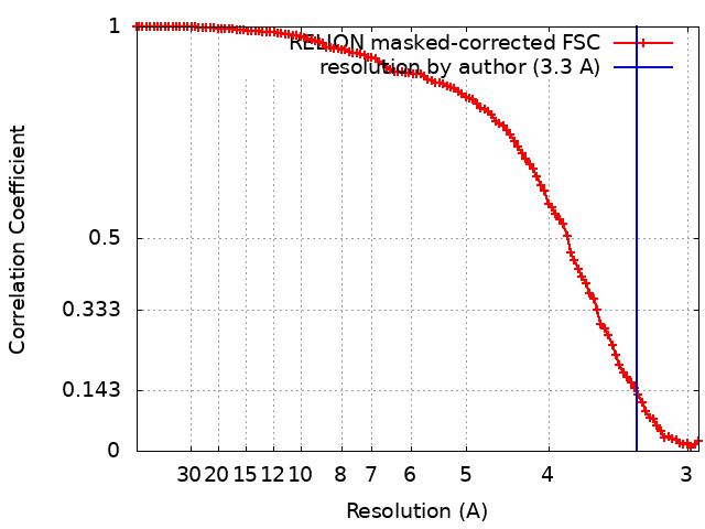
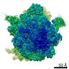
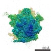
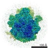
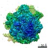
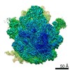
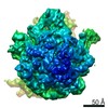
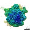
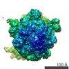
Journal: RNA / Year: 2022 Title: The landscape of translational stall sites in bacteria revealed by monosome and disome profiling. Authors: Tomoya Fujita / Takeshi Yokoyama / Mikako Shirouzu / Hideki Taguchi / Takuhiro Ito / Shintaro Iwasaki / Abstract: Ribosome pauses are associated with various cotranslational events and determine the fate of mRNAs and proteins. Thus, the identification of precise pause sites across the transcriptome is desirable; ...Ribosome pauses are associated with various cotranslational events and determine the fate of mRNAs and proteins. Thus, the identification of precise pause sites across the transcriptome is desirable; however, the landscape of ribosome pauses in bacteria remains ambiguous. Here, we harness monosome and disome (or collided ribosome) profiling strategies to survey ribosome pause sites in Compared to eukaryotes, ribosome collisions in bacteria showed remarkable differences: a low frequency of disomes at stop codons, collisions occurring immediately after 70S assembly on start codons, and shorter queues of ribosomes trailing upstream. The pause sites corresponded with the biochemical validation by integrated nascent chain profiling (iNP) to detect polypeptidyl-tRNA, an elongation intermediate. Moreover, the subset of those sites showed puromycin resistance, presenting slow peptidyl transfer. Among the identified sites, the ribosome pause at Asn586 of was validated by biochemical reporter assay, tRNA sequencing (tRNA-seq), and cryo-electron microscopy (cryo-EM) experiments. Our results provide a useful resource for ribosome stalling sites in bacteria.
History
Deposition
Aug 7, 2020
-
Header (metadata) release
Dec 8, 2021
-
Map release
Dec 8, 2021
-
Update
Jul 1, 2026
-
Current status
Jul 1, 2026
Processing site: PDBj / Status: Released
-
Structure visualization
Movie
Surface view with section colored by density value
In the structure databanks used in Yorodumi, some data are registered as the other names, "COVID-19 virus" and "2019-nCoV". Here are the details of the virus and the list of structure data.
Jan 31, 2019. EMDB accession codes are about to change! (news from PDBe EMDB page)
EMDB accession codes are about to change! (news from PDBe EMDB page)
The allocation of 4 digits for EMDB accession codes will soon come to an end. Whilst these codes will remain in use, new EMDB accession codes will include an additional digit and will expand incrementally as the available range of codes is exhausted. The current 4-digit format prefixed with “EMD-” (i.e. EMD-XXXX) will advance to a 5-digit format (i.e. EMD-XXXXX), and so on. It is currently estimated that the 4-digit codes will be depleted around Spring 2019, at which point the 5-digit format will come into force.
The EM Navigator/Yorodumi systems omit the EMD- prefix.
Related info.:Q: What is EMD? / ID/Accession-code notation in Yorodumi/EM Navigator
Yorodumi is a browser for structure data from EMDB, PDB, SASBDB, etc.
This page is also the successor to EM Navigator detail page, and also detail information page/front-end page for Omokage search.
The word "yorodu" (or yorozu) is an old Japanese word meaning "ten thousand". "mi" (miru) is to see.
Related info.:EMDB / PDB / SASBDB / Comparison of 3 databanks / Yorodumi Search / Aug 31, 2016. New EM Navigator & Yorodumi / Yorodumi Papers / Jmol/JSmol / Function and homology information / Changes in new EM Navigator and Yorodumi
 Movie
Movie Controller


 Open data
 Basic information
 Map data
 Sample
 Keywords
 Function and homology information
 Authors
 Japan, 1 items
  Citation
 Structure visualization
 Downloads & links
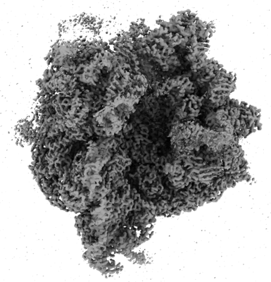 emd_30431.png
 http://ftp.pdbj.org/pub/emdb/structures/EMD-30431
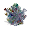


 Z (Sec.)
 Y (Row.)
 X (Col.)


 Sample components

 Processing
 Electron microscopy
 FIELD EMISSION GUN