 Movie
Movie Controller

[English] 日本語
 Yorodumi
- EMDB-30016: Cryo-EM structure of the calcium homeostasis modulator 1 channel -
+ Open data

- Basic information
| Entry | Database: EMDB / ID: EMD-30016 | ||||||||||||||||||
|---|---|---|---|---|---|---|---|---|---|---|---|---|---|---|---|---|---|---|---|
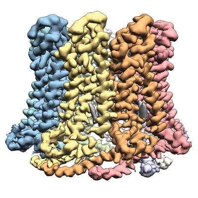
| Title | Cryo-EM structure of the calcium homeostasis modulator 1 channel | ||||||||||||||||||
 Map data Map data | |||||||||||||||||||
 Sample Sample |
| ||||||||||||||||||
| Function / homology | Calcium homeostasis modulator family / Calcium homeostasis modulator / voltage-gated calcium channel activity / monoatomic cation channel activity / plasma membrane / Calcium homeostasis modulator 1 Function and homology information Function and homology information | ||||||||||||||||||
| Biological species |  | ||||||||||||||||||
| Method | single particle reconstruction / cryo EM / Resolution: 3.1 Å | ||||||||||||||||||
 Authors Authors | Ren Y / Yang X / Shen Y | ||||||||||||||||||
| Funding support |  China, 5 items China, 5 items
| ||||||||||||||||||
 Citation Citation | Journal: Sci Adv / Year: 2020 Title: Cryo-EM structure of the calcium homeostasis modulator 1 channel. Authors: Yue Ren / Tianlei Wen / Zhiqin Xi / Shunjin Li / Jing Lu / Xing Zhang / Xue Yang / Yuequan Shen / Abstract: Calcium homeostasis modulator 1 (CALHM1) is a voltage-gated ATP release channel that plays an important role in neural gustatory signaling and the pathogenesis of Alzheimer's disease. Here, we ...Calcium homeostasis modulator 1 (CALHM1) is a voltage-gated ATP release channel that plays an important role in neural gustatory signaling and the pathogenesis of Alzheimer's disease. Here, we present a cryo-electron microscopy structure of full-length Ca-free CALHM1 from Danio rerio at an overall resolution of 3.1 Å. Our structure reveals an octameric architecture with a wide pore diameter of ~20 Å, presumably representing the active conformation. The overall structure is substantially different from that of the isoform CALHM2, which forms both undecameric hemichannels and gap junctions. The N-terminal small helix folds back to the pore and forms an antiparallel interaction with transmembrane helix 1. Structural analysis revealed that the extracellular loop 1 region within the dimer interface may contribute to oligomeric assembly. A positive potential belt inside the pore was identified that may modulate ion permeation. Our structure offers insights into the assembly and gating mechanism of the CALHM1 channel. | ||||||||||||||||||
| History |
|
- Structure visualization
| Movie |
Movie viewer |
|---|---|
| Structure viewer | EM map: SurfViewMolmilJmol/JSmol |
| Supplemental images |
- Downloads & links
-EMDB archive
| Map data | emd_30016.map.gz | 5.8 MB | EMDB map data format | |
|---|---|---|---|---|
| Header (meta data) | emd-30016-v30.xmlemd-30016.xml | 10.9 KB 10.9 KB | Display Display | EMDB header |
| Images |  emd_30016.png emd_30016.png | 220.8 KB | ||
| Archive directory |  http://ftp.pdbj.org/pub/emdb/structures/EMD-30016ftp://ftp.pdbj.org/pub/emdb/structures/EMD-30016 http://ftp.pdbj.org/pub/emdb/structures/EMD-30016ftp://ftp.pdbj.org/pub/emdb/structures/EMD-30016 | HTTPS FTP |
-Validation report
| Summary document | emd_30016_validation.pdf.gz | 350 KB | Display | EMDB validaton report |
|---|---|---|---|---|
| Full document | emd_30016_full_validation.pdf.gz | 349.6 KB | Display | |
| Data in XML | emd_30016_validation.xml.gz | 6.1 KB | Display | |
| Data in CIF | emd_30016_validation.cif.gz | 7 KB | Display | |
| Arichive directory | https://ftp.pdbj.org/pub/emdb/validation_reports/EMD-30016ftp://ftp.pdbj.org/pub/emdb/validation_reports/EMD-30016 | HTTPS FTP |
-Related structure data
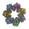
| Related structure data |  6lygMC M: atomic model generated by this map C: citing same article ( |
|---|---|
| Similar structure data |


-Links
| EMDB pages | EMDB (EBI/PDBe) / EMDataResource |
|---|
-Map
| File | Download / File: emd_30016.map.gz / Format: CCP4 / Size: 67 MB / Type: IMAGE STORED AS FLOATING POINT NUMBER (4 BYTES) | ||||||||||||||||||||||||||||||||||||||||||||||||||||||||||||||||||||
|---|---|---|---|---|---|---|---|---|---|---|---|---|---|---|---|---|---|---|---|---|---|---|---|---|---|---|---|---|---|---|---|---|---|---|---|---|---|---|---|---|---|---|---|---|---|---|---|---|---|---|---|---|---|---|---|---|---|---|---|---|---|---|---|---|---|---|---|---|---|
| Voxel size | X=Y=Z: 1.014 Å | ||||||||||||||||||||||||||||||||||||||||||||||||||||||||||||||||||||
| Density |
| ||||||||||||||||||||||||||||||||||||||||||||||||||||||||||||||||||||
| Symmetry | Space group: 1 | ||||||||||||||||||||||||||||||||||||||||||||||||||||||||||||||||||||
| Details | EMDB XML:
CCP4 map header:
| ||||||||||||||||||||||||||||||||||||||||||||||||||||||||||||||||||||
-Supplemental data
- Sample components
-Entire : CALHM1 channel
| Entire | Name: CALHM1 channel |
|---|---|
| Components |
|
-Supramolecule #1: CALHM1 channel
| Supramolecule | Name: CALHM1 channel / type: cell / ID: 1 / Parent: 0 / Macromolecule list: #1 |
|---|---|
| Source (natural) | Organism: |
-Macromolecule #1: Calcium homeostasis modulator 1
| Macromolecule | Name: Calcium homeostasis modulator 1 / type: protein_or_peptide / ID: 1 / Number of copies: 8 / Enantiomer: LEVO |
|---|---|
| Source (natural) | Organism: |
| Molecular weight | Theoretical: 41.213645 KDa |
| Recombinant expression | Organism:  Homo sapiens (human) Homo sapiens (human) |
| Sequence | String: LEQKLISEED LRSDKFRIMV QFLQANQESF MNGICGIMAL ASAQMYSSFE FTCPCLPDYN YAYGIGILIV PPIWFFLLGY VMNNNISVL TEEWKRPVGK RSKDPAVLRY MFSSMTQRAL IAPAVWIAVT LMDGKSFLCA FSPTADLSEF VNESYQSLSQ K ELLKIQAK ...String: LEQKLISEED LRSDKFRIMV QFLQANQESF MNGICGIMAL ASAQMYSSFE FTCPCLPDYN YAYGIGILIV PPIWFFLLGY VMNNNISVL TEEWKRPVGK RSKDPAVLRY MFSSMTQRAL IAPAVWIAVT LMDGKSFLCA FSPTADLSEF VNESYQSLSQ K ELLKIQAK IPCKDIFEEH EIISREAATR YIRCLSQACG WTFLMVITLV AFLVRAIRPC FTQAAFLKTK YWSHYIDTER KL FDETCKE HAKSFAKVCI QQYFESISGE IVSQLPQSPA KKGKGNKDED GEKQKSDEER LLGIRKEGDM NKVLWNWHTC KPP LLLSKR TEEMNGHAHL DTHSLTDERH TKKKAVVYYS KV |
-Macromolecule #2: 2-acetamido-2-deoxy-beta-D-glucopyranose
| Macromolecule | Name: 2-acetamido-2-deoxy-beta-D-glucopyranose / type: ligand / ID: 2 / Number of copies: 8 / Formula: NAG |
|---|---|
| Molecular weight | Theoretical: 221.208 Da |
| Chemical component information |  ChemComp-NAG: |
-Experimental details
-Structure determination
| Method | cryo EM |
|---|---|
 Processing Processing | single particle reconstruction |
| Aggregation state | particle |
-Sample preparation
| Concentration | 8 mg/mL |
|---|---|
| Buffer | pH: 7.5 |
| Vitrification | Cryogen name: ETHANE / Instrument: FEI VITROBOT MARK IV |
- Electron microscopy
| Microscope | FEI TITAN KRIOS |
|---|---|
| Image recording | Film or detector model: GATAN K2 SUMMIT (4k x 4k) / Detector mode: COUNTING / Average electron dose: 50.0 e/Å2 |
| Electron beam | Acceleration voltage: 300 kV / Electron source:  FIELD EMISSION GUN FIELD EMISSION GUN |
| Electron optics | Illumination mode: FLOOD BEAM / Imaging mode: BRIGHT FIELD |
| Sample stage | Specimen holder model: FEI TITAN KRIOS AUTOGRID HOLDER / Cooling holder cryogen: NITROGEN |
| Experimental equipment |  Model: Titan Krios / Image courtesy: FEI Company |
-Image processing
| Startup model | Type of model: OTHER / Details: Ab initial by Cryosparc2 |
|---|---|
| Final reconstruction | Resolution.type: BY AUTHOR / Resolution: 3.1 Å / Resolution method: FSC 0.143 CUT-OFF / Number images used: 59833 |
| Initial angle assignment | Type: NOT APPLICABLE |
| Final angle assignment | Type: NOT APPLICABLE |
