 Movie
Movie Controller

[English] 日本語
 Yorodumi
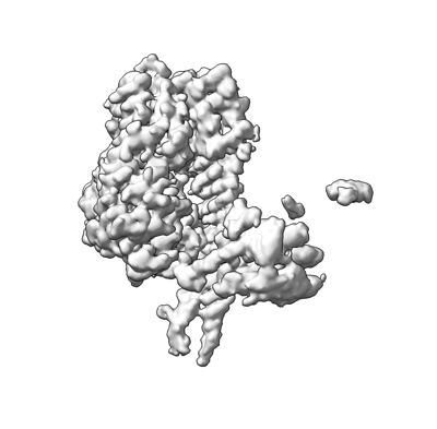
- EMDB-29913: Cryo-EM 3D map of the Mycobacterium tuberculosis Hsp70 protein Dn... -
+ Open data

- Basic information
| Entry |  | |||||||||
|---|---|---|---|---|---|---|---|---|---|---|
| Title | Cryo-EM 3D map of the Mycobacterium tuberculosis Hsp70 protein DnaK bound to the nucleotide exchange factor GrpE | |||||||||
 Map data Map data | processed by DeepEMhancer | |||||||||
 Sample Sample |
| |||||||||
 Keywords Keywords | heat shock protein 70 / nucleotide exchange factor / protein folding and refolding / CHAPERONE | |||||||||
| Biological species |   Mycobacterium tuberculosis (bacteria) Mycobacterium tuberculosis (bacteria) | |||||||||
| Method | single particle reconstruction / cryo EM / Resolution: 3.7 Å | |||||||||
 Authors Authors | Xiao X / Li H | |||||||||
| Funding support |  United States, 1 items United States, 1 items
| |||||||||
 Citation Citation | Journal: Nat Commun / Year: 2024 Title: Structure of the M. tuberculosis DnaK-GrpE complex reveals how key DnaK roles are controlled. Authors: Xiansha Xiao / Allison Fay / Pablo Santos Molina / Amanda Kovach / Michael S Glickman / Huilin Li / Abstract: The molecular chaperone DnaK is essential for viability of Mycobacterium tuberculosis (Mtb). DnaK hydrolyzes ATP to fold substrates, and the resulting ADP is exchanged for ATP by the nucleotide ...The molecular chaperone DnaK is essential for viability of Mycobacterium tuberculosis (Mtb). DnaK hydrolyzes ATP to fold substrates, and the resulting ADP is exchanged for ATP by the nucleotide exchange factor GrpE. It has been unclear how GrpE couples DnaK's nucleotide exchange with substrate release. Here we report a cryo-EM analysis of GrpE bound to an intact Mtb DnaK, revealing an asymmetric 1:2 DnaK-GrpE complex. The GrpE dimer ratchets to modulate both DnaK nucleotide-binding domain and the substrate-binding domain. We further show that the disordered GrpE N-terminus is critical for substrate release, and that the DnaK-GrpE interface is essential for protein folding activity both in vitro and in vivo. Therefore, the Mtb GrpE dimer allosterically regulates DnaK to concomitantly release ADP in the nucleotide-binding domain and substrate peptide in the substrate-binding domain. | |||||||||
| History |
|
- Structure visualization
| Supplemental images |
|---|
- Downloads & links
-EMDB archive
| Map data | emd_29913.map.gz | 109.7 MB |  EMDB map data format EMDB map data format | |
|---|---|---|---|---|
| Header (meta data) | emd-29913-v30.xmlemd-29913.xml | 15.3 KB 15.3 KB | Display Display | EMDB header |
| Images |  emd_29913.png emd_29913.png | 66.6 KB | ||
| Filedesc metadata | emd-29913.cif.gz | 4.5 KB | ||
| Others | emd_29913_half_map_1.map.gzemd_29913_half_map_2.map.gz | 116.2 MB 116.2 MB | ||
| Archive directory |  http://ftp.pdbj.org/pub/emdb/structures/EMD-29913ftp://ftp.pdbj.org/pub/emdb/structures/EMD-29913 http://ftp.pdbj.org/pub/emdb/structures/EMD-29913ftp://ftp.pdbj.org/pub/emdb/structures/EMD-29913 | HTTPS FTP |
-Validation report
| Summary document | emd_29913_validation.pdf.gz | 698.3 KB | Display | EMDB validaton report |
|---|---|---|---|---|
| Full document | emd_29913_full_validation.pdf.gz | 697.9 KB | Display | |
| Data in XML | emd_29913_validation.xml.gz | 13.8 KB | Display | |
| Data in CIF | emd_29913_validation.cif.gz | 16.3 KB | Display | |
| Arichive directory | https://ftp.pdbj.org/pub/emdb/validation_reports/EMD-29913ftp://ftp.pdbj.org/pub/emdb/validation_reports/EMD-29913 | HTTPS FTP |
-Related structure data


-Links
| EMDB pages | EMDB (EBI/PDBe) / EMDataResource |
|---|
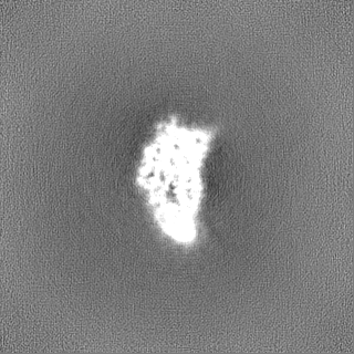
-Map
| File | Download / File: emd_29913.map.gz / Format: CCP4 / Size: 125 MB / Type: IMAGE STORED AS FLOATING POINT NUMBER (4 BYTES) | ||||||||||||||||||||
|---|---|---|---|---|---|---|---|---|---|---|---|---|---|---|---|---|---|---|---|---|---|
| Annotation | processed by DeepEMhancer | ||||||||||||||||||||
| Voxel size | X=Y=Z: 0.828 Å | ||||||||||||||||||||
| Density |
| ||||||||||||||||||||
| Symmetry | Space group: 1 | ||||||||||||||||||||
| Details | EMDB XML:
|
-Supplemental data
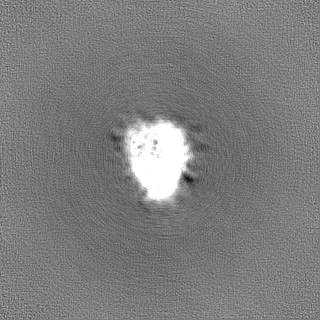
-Half map: #1
| File | emd_29913_half_map_1.map | ||||||||||||
|---|---|---|---|---|---|---|---|---|---|---|---|---|---|
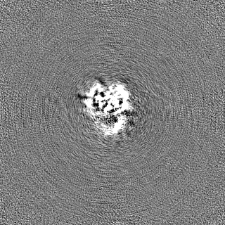
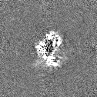
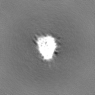
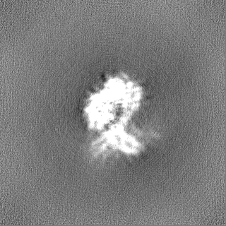
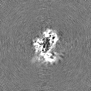
| Projections & Slices |
| ||||||||||||
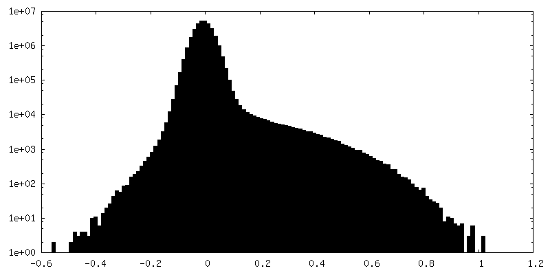
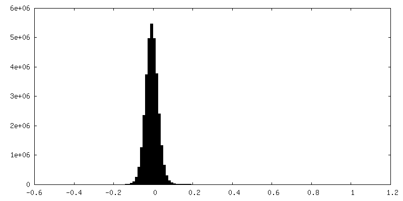
| Density Histograms |
 Z
Z Y
Y X
X
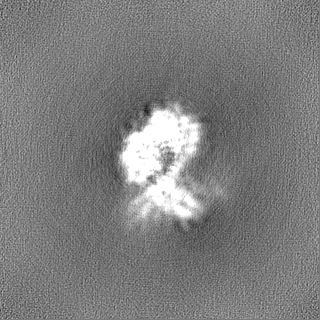


-Half map: #2
| File | emd_29913_half_map_2.map | ||||||||||||
|---|---|---|---|---|---|---|---|---|---|---|---|---|---|
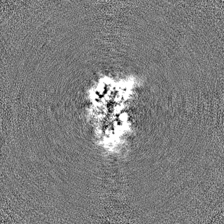
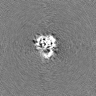
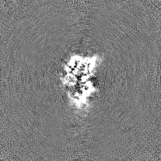
| Projections & Slices |
| ||||||||||||
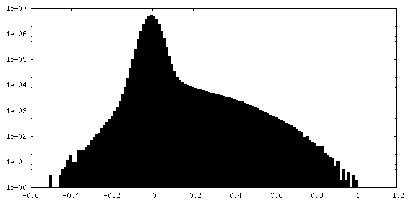
| Density Histograms |

- Sample components
-Entire : Binary complex of DnaK with nucleotide exchange factor GrpE
| Entire | Name: Binary complex of DnaK with nucleotide exchange factor GrpE |
|---|---|
| Components |
|
-Supramolecule #1: Binary complex of DnaK with nucleotide exchange factor GrpE
| Supramolecule | Name: Binary complex of DnaK with nucleotide exchange factor GrpE type: complex / ID: 1 / Parent: 0 / Macromolecule list: #1-#2 |
|---|---|
| Source (natural) | Organism: Mycobacterium tuberculosis (bacteria) |
-Experimental details
-Structure determination
| Method | cryo EM |
|---|---|
 Processing Processing | single particle reconstruction |
| Aggregation state | particle |
-Sample preparation
| Buffer | pH: 8 |
|---|---|
| Grid | Model: Quantifoil R2/1 / Support film - Material: CARBON |
| Vitrification | Cryogen name: ETHANE / Chamber humidity: 100 % / Chamber temperature: 283.15 K / Instrument: FEI VITROBOT MARK II |
- Electron microscopy
| Microscope | FEI TITAN KRIOS |
|---|---|
| Specialist optics | Energy filter - Name: GIF Bioquantum / Energy filter - Slit width: 20 eV |
| Image recording | Film or detector model: GATAN K3 BIOQUANTUM (6k x 4k) / Detector mode: SUPER-RESOLUTION / Number real images: 15720 / Average exposure time: 1.5 sec. / Average electron dose: 66.0 e/Å2 |
| Electron beam | Acceleration voltage: 300 kV / Electron source:  FIELD EMISSION GUN FIELD EMISSION GUN |
| Electron optics | C2 aperture diameter: 100.0 µm / Illumination mode: FLOOD BEAM / Imaging mode: BRIGHT FIELD / Cs: 2.7 mm / Nominal defocus max: 1.8 µm / Nominal defocus min: 1.3 µm / Nominal magnification: 105000 |
| Sample stage | Specimen holder model: FEI TITAN KRIOS AUTOGRID HOLDER / Cooling holder cryogen: NITROGEN |
| Experimental equipment |  Model: Titan Krios / Image courtesy: FEI Company |

