 ムービー
ムービー コントローラー


+ データを開く

- 基本情報
基本情報
| 登録情報 | 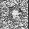 | |||||||||
|---|---|---|---|---|---|---|---|---|---|---|
| タイトル | IMC surface filament, sawtooth conformation, from Cryptosporidium parvum sporozoites | |||||||||
 マップデータ マップデータ | Subtomogram average of the IMC surface filaments, sawtooth conformation, from Cryptosporidium parvum sporozoites | |||||||||
 試料 試料 |
| |||||||||
 キーワード キーワード | Parasite / Inner membrane complex / apicomplexa / CELL INVASION | |||||||||
| 生物種 |  Cryptosporidium parvum Iowa (小形クリプトスポリジウム) Cryptosporidium parvum Iowa (小形クリプトスポリジウム) | |||||||||
| 手法 | サブトモグラム平均法 / クライオ電子顕微鏡法 / 解像度: 47.0 Å | |||||||||
 データ登録者 データ登録者 | Martinez M / Mageswaran SK / Chang Y-W | |||||||||
| 資金援助 |  米国, 1件 米国, 1件
| |||||||||
 引用 引用 | ジャーナル: Nat Commun / 年: 2023 タイトル: Origin and arrangement of actin filaments for gliding motility in apicomplexan parasites revealed by cryo-electron tomography. 著者: Matthew Martinez / Shrawan Kumar Mageswaran / Amandine Guérin / William David Chen / Cameron Parker Thompson / Sabine Chavin / Dominique Soldati-Favre / Boris Striepen / Yi-Wei Chang /  要旨: The phylum Apicomplexa comprises important eukaryotic parasites that invade host tissues and cells using a unique mechanism of gliding motility. Gliding is powered by actomyosin motors that ...The phylum Apicomplexa comprises important eukaryotic parasites that invade host tissues and cells using a unique mechanism of gliding motility. Gliding is powered by actomyosin motors that translocate host-attached surface adhesins along the parasite cell body. Actin filaments (F-actin) generated by Formin1 play a central role in this critical parasitic activity. However, their subcellular origin, path and ultrastructural arrangement are poorly understood. Here we used cryo-electron tomography to image motile Cryptosporidium parvum sporozoites and reveal the cellular architecture of F-actin at nanometer-scale resolution. We demonstrate that F-actin nucleates at the apically positioned preconoidal rings and is channeled into the pellicular space between the parasite plasma membrane and the inner membrane complex in a conoid extrusion-dependent manner. Within the pellicular space, filaments on the inner membrane complex surface appear to guide the apico-basal flux of F-actin. F-actin concordantly accumulates at the basal end of the parasite. Finally, analyzing a Formin1-depleted Toxoplasma gondii mutant pinpoints the upper preconoidal ring as the conserved nucleation hub for F-actin in Cryptosporidium and Toxoplasma. Together, we provide an ultrastructural model for the life cycle of F-actin for apicomplexan gliding motility. | |||||||||
| 履歴 |
|
- 構造の表示
構造の表示
| 添付画像 |
|---|

- ダウンロードとリンク
-EMDBアーカイブ
| マップデータ | emd_29809.map.gz | 14.5 MB |  EMDBマップデータ形式 EMDBマップデータ形式 | |
|---|---|---|---|---|
| ヘッダ (付随情報) | emd-29809-v30.xmlemd-29809.xml | 15.6 KB 15.6 KB | 表示 表示 | EMDBヘッダ |
| FSC (解像度算出) | emd_29809_fsc.xml | 5.7 KB | 表示 | FSCデータファイル |
| 画像 | 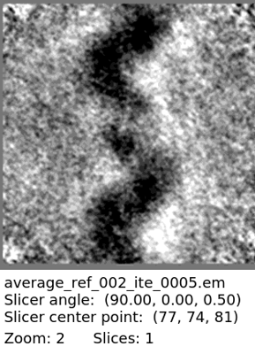 emd_29809.png emd_29809.png | 98.2 KB | ||
| マスクデータ | emd_29809_msk_1.map | 15.6 MB | マスクマップ | |
| その他 | emd_29809_half_map_1.map.gzemd_29809_half_map_2.map.gz | 14.5 MB 14.5 MB | ||
| アーカイブディレクトリ |  http://ftp.pdbj.org/pub/emdb/structures/EMD-29809ftp://ftp.pdbj.org/pub/emdb/structures/EMD-29809 http://ftp.pdbj.org/pub/emdb/structures/EMD-29809ftp://ftp.pdbj.org/pub/emdb/structures/EMD-29809 | HTTPS FTP |
-検証レポート
| 文書・要旨 | emd_29809_validation.pdf.gz | 1.2 MB | 表示 | EMDB検証レポート |
|---|---|---|---|---|
| 文書・詳細版 | emd_29809_full_validation.pdf.gz | 1.2 MB | 表示 | |
| XML形式データ | emd_29809_validation.xml.gz | 11.3 KB | 表示 | |
| CIF形式データ | emd_29809_validation.cif.gz | 15.5 KB | 表示 | |
| アーカイブディレクトリ | https://ftp.pdbj.org/pub/emdb/validation_reports/EMD-29809ftp://ftp.pdbj.org/pub/emdb/validation_reports/EMD-29809 | HTTPS FTP |
-関連構造データ
| 関連構造データ | C: 同じ文献を引用 ( |
|---|


-リンク
| EMDBのページ | EMDB (EBI/PDBe) / EMDataResource |
|---|
-マップ
| ファイル | ダウンロード / ファイル: emd_29809.map.gz / 形式: CCP4 / 大きさ: 15.6 MB / タイプ: IMAGE STORED AS FLOATING POINT NUMBER (4 BYTES) | ||||||||||||||||||||||||||||||||||||
|---|---|---|---|---|---|---|---|---|---|---|---|---|---|---|---|---|---|---|---|---|---|---|---|---|---|---|---|---|---|---|---|---|---|---|---|---|---|
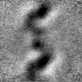
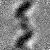
| 注釈 | Subtomogram average of the IMC surface filaments, sawtooth conformation, from Cryptosporidium parvum sporozoites | ||||||||||||||||||||||||||||||||||||
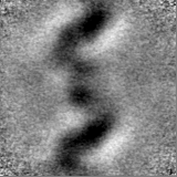
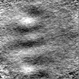
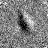
| 投影像・断面図 | 画像のコントロール
画像は Spider により作成 | ||||||||||||||||||||||||||||||||||||
| ボクセルのサイズ | X=Y=Z: 2.65 Å | ||||||||||||||||||||||||||||||||||||
| 密度 |
| ||||||||||||||||||||||||||||||||||||
| 対称性 | 空間群: 1 | ||||||||||||||||||||||||||||||||||||
| 詳細 | EMDB XML:
|
 Z (Sec.)
 Y (Row.)
 X (Col.)


-添付データ
-マスク #1
| ファイル | emd_29809_msk_1.map | ||||||||||||
|---|---|---|---|---|---|---|---|---|---|---|---|---|---|
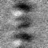
| 投影像・断面図 |
| ||||||||||||
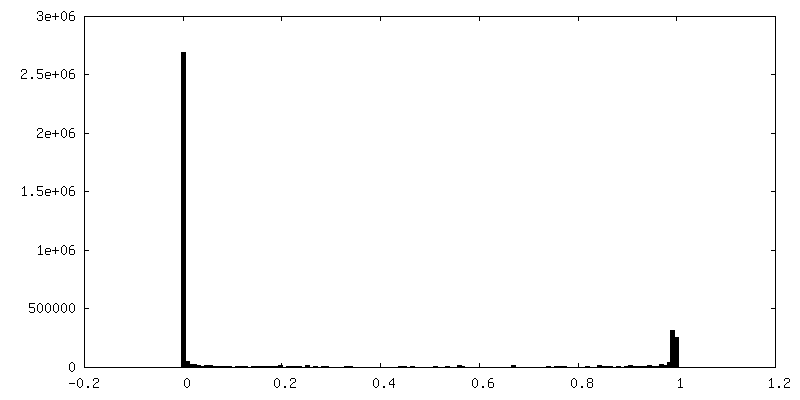
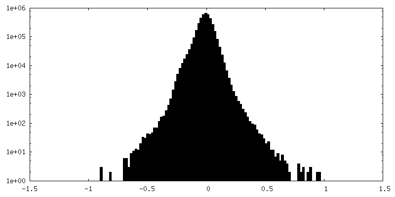
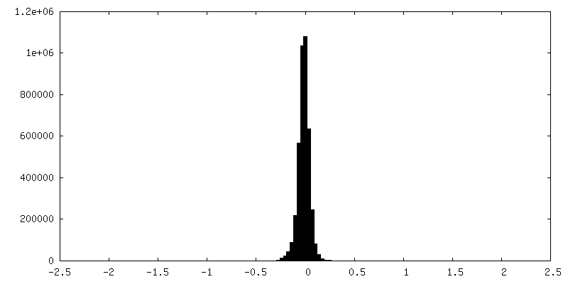
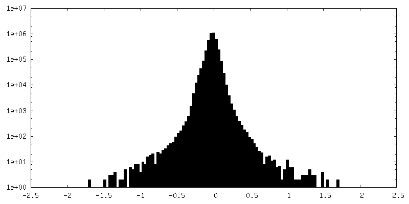
| 密度ヒストグラム |


-ハーフマップ: #1
| ファイル | emd_29809_half_map_1.map | ||||||||||||
|---|---|---|---|---|---|---|---|---|---|---|---|---|---|
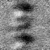
| 投影像・断面図 |
| ||||||||||||
| 密度ヒストグラム |


-ハーフマップ: #2
| ファイル | emd_29809_half_map_2.map | ||||||||||||
|---|---|---|---|---|---|---|---|---|---|---|---|---|---|
| 投影像・断面図 |
| ||||||||||||
| 密度ヒストグラム |

- 試料の構成要素
-全体 : IMC surface filaments, sawtooth conformation
| 全体 | 名称: IMC surface filaments, sawtooth conformation |
|---|---|
| 要素 |
|
-超分子 #1: IMC surface filaments, sawtooth conformation
| 超分子 | 名称: IMC surface filaments, sawtooth conformation / タイプ: organelle_or_cellular_component / ID: 1 / 親要素: 0 詳細: In situ structure of the IMC surface filaments, sawtooth conformation, from Cryptosporidium parvum sporozoites |
|---|---|
| 由来(天然) | 生物種: Cryptosporidium parvum Iowa (小形クリプトスポリジウム) |
-実験情報
-構造解析
| 手法 | クライオ電子顕微鏡法 |
|---|---|
 解析 解析 | サブトモグラム平均法 |
| 試料の集合状態 | particle |
-試料調製
| 緩衝液 | pH: 7.4 |
|---|---|
| グリッド | モデル: Quantifoil R2/2 / 材質: COPPER / メッシュ: 200 / 支持フィルム - 材質: CARBON / 支持フィルム - トポロジー: HOLEY / 前処理 - タイプ: GLOW DISCHARGE |
| 凍結 | 凍結剤: ETHANE-PROPANE / チャンバー内湿度: 99 % / チャンバー内温度: 310 K / 装置: LEICA EM GP 詳細: Isolated sporozoites were resuspended in media, and 4 uL was applied to the carbon side of the grid and front blotted for 4s. |
| 詳細 | Sample was averaged from within frozen-hydrated Cryptosporidium parvum sporozoites |
- 電子顕微鏡法
| 顕微鏡 | FEI TITAN KRIOS |
|---|---|
| 特殊光学系 | 位相板: VOLTA PHASE PLATE / エネルギーフィルター - 名称: GIF Bioquantum / エネルギーフィルター - スリット幅: 20 eV |
| 撮影 | フィルム・検出器のモデル: GATAN K3 (6k x 4k) / 平均露光時間: 0.4 sec. / 平均電子線量: 2.3 e/Å2 |
| 電子線 | 加速電圧: 300 kV / 電子線源:  FIELD EMISSION GUN FIELD EMISSION GUN |
| 電子光学系 | 照射モード: FLOOD BEAM / 撮影モード: BRIGHT FIELD / 最大 デフォーカス(公称値): 4.0 µm / 最小 デフォーカス(公称値): 1.5 µm / 倍率(公称値): 33000 |
| 試料ステージ | 試料ホルダーモデル: FEI TITAN KRIOS AUTOGRID HOLDER |
| 実験機器 |  モデル: Titan Krios / 画像提供: FEI Company |
-画像解析
| 最終 再構成 | 想定した対称性 - 点群: C1 (非対称) / 解像度のタイプ: BY AUTHOR / 解像度: 47.0 Å / 解像度の算出法: FSC 0.5 CUT-OFF / ソフトウェア - 名称: Dynamo (ver. 1.1.509) / 使用したサブトモグラム数: 929 |
|---|---|
| 抽出 | トモグラム数: 24 / 使用した粒子像数: 6709 / 手法: Filaments traced / ソフトウェア - 名称: Dynamo (ver. 1.1.509) 詳細: Traced filaments in IMOD. Imported coordinates into Dynamo, where points were placed along each filament trace. |
| 最終 角度割当 | タイプ: NOT APPLICABLE / ソフトウェア - 名称: Dynamo (ver. 1.1.509) |
| FSC曲線 (解像度の算出) |  |
