 Movie
Movie Controller

[English] 日本語
 Yorodumi
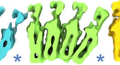
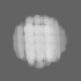
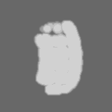
- EMDB-29442: Subtomogram of the 4-beta-ring hoop of M. hungatei sheath structure. -
+ Open data

- Basic information
| Entry |  | |||||||||||||||
|---|---|---|---|---|---|---|---|---|---|---|---|---|---|---|---|---|
| Title | Subtomogram of the 4-beta-ring hoop of M. hungatei sheath structure. | |||||||||||||||
 Map data Map data | sub-tomogram average of 4-beta-ring hoop | |||||||||||||||
 Sample Sample |
| |||||||||||||||
 Keywords Keywords | M. hungatei / archaeal sheath / cell wall / protein translocation / sheath assembly / cryoET / subtomogram average / amyloid-like protein / cell growth / STRUCTURAL PROTEIN | |||||||||||||||
| Biological species |  Methanospirillum hungatei JF-1 (archaea) Methanospirillum hungatei JF-1 (archaea) | |||||||||||||||
| Method | subtomogram averaging / cryo EM / Resolution: 7.92 Å | |||||||||||||||
 Authors Authors | Wang H / Zhang J / Zhou ZH | |||||||||||||||
| Funding support |  United States, 4 items United States, 4 items
| |||||||||||||||
 Citation Citation | Journal: Nat Commun / Year: 2023 Title: Hierarchical organization and assembly of the archaeal cell sheath from an amyloid-like protein. Authors: Hui Wang / Jiayan Zhang / Daniel Toso / Shiqing Liao / Farzaneh Sedighian / Robert Gunsalus / Z Hong Zhou / Abstract: Certain archaeal cells possess external proteinaceous sheath, whose structure and organization are both unknown. By cellular cryogenic electron tomography (cryoET), here we have determined sheath ...Certain archaeal cells possess external proteinaceous sheath, whose structure and organization are both unknown. By cellular cryogenic electron tomography (cryoET), here we have determined sheath organization of the prototypical archaeon, Methanospirillum hungatei. Fitting of Alphafold-predicted model of the sheath protein (SH) monomer into the 7.9 Å-resolution structure reveals that the sheath cylinder consists of axially stacked β-hoops, each of which is comprised of two to six 400 nm-diameter rings of β-strand arches (β-rings). With both similarities to and differences from amyloid cross-β fibril architecture, each β-ring contains two giant β-sheets contributed by ~ 450 SH monomers that entirely encircle the outer circumference of the cell. Tomograms of immature cells suggest models of sheath biogenesis: oligomerization of SH monomers into β-ring precursors after their membrane-proximal cytoplasmic synthesis, followed by translocation through the unplugged end of a dividing cell, and insertion of nascent β-hoops into the immature sheath cylinder at the junction of two daughter cells. | |||||||||||||||
| History |
|
- Structure visualization
| Supplemental images |
|---|
- Downloads & links
-EMDB archive
| Map data | emd_29442.map.gz | 11.9 MB |  EMDB map data format EMDB map data format | |
|---|---|---|---|---|
| Header (meta data) | emd-29442-v30.xmlemd-29442.xml | 14 KB 14 KB | Display Display | EMDB header |
| Images |  emd_29442.png emd_29442.png | 83.1 KB | ||
| Masks | emd_29442_msk_1.map | 15.6 MB | Mask map | |
| Filedesc metadata | emd-29442.cif.gz | 4.1 KB | ||
| Others | emd_29442_half_map_1.map.gzemd_29442_half_map_2.map.gz | 7.7 MB 7.7 MB | ||
| Archive directory |  http://ftp.pdbj.org/pub/emdb/structures/EMD-29442ftp://ftp.pdbj.org/pub/emdb/structures/EMD-29442 http://ftp.pdbj.org/pub/emdb/structures/EMD-29442ftp://ftp.pdbj.org/pub/emdb/structures/EMD-29442 | HTTPS FTP |
-Related structure data


-Links
| EMDB pages | EMDB (EBI/PDBe) / EMDataResource |
|---|
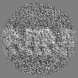
-Map
| File | Download / File: emd_29442.map.gz / Format: CCP4 / Size: 15.6 MB / Type: IMAGE STORED AS FLOATING POINT NUMBER (4 BYTES) | ||||||||||||||||||||||||||||||||||||
|---|---|---|---|---|---|---|---|---|---|---|---|---|---|---|---|---|---|---|---|---|---|---|---|---|---|---|---|---|---|---|---|---|---|---|---|---|---|
| Annotation | sub-tomogram average of 4-beta-ring hoop | ||||||||||||||||||||||||||||||||||||
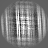
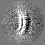
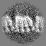
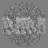
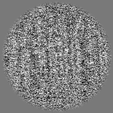
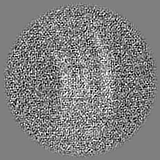
| Projections & slices | Image control
Images are generated by Spider. | ||||||||||||||||||||||||||||||||||||
| Voxel size | X=Y=Z: 1.634 Å | ||||||||||||||||||||||||||||||||||||
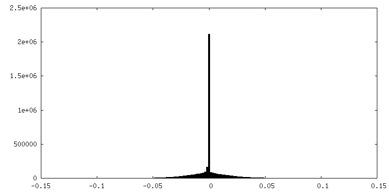
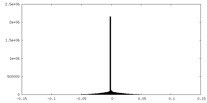
| Density |
| ||||||||||||||||||||||||||||||||||||
| Symmetry | Space group: 1 | ||||||||||||||||||||||||||||||||||||
| Details | EMDB XML:
|
 Z (Sec.)
 Y (Row.)
 X (Col.)


-Supplemental data
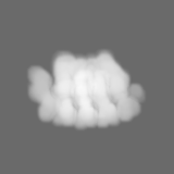
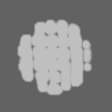
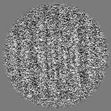
-Mask #1
| File | emd_29442_msk_1.map | ||||||||||||
|---|---|---|---|---|---|---|---|---|---|---|---|---|---|
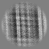
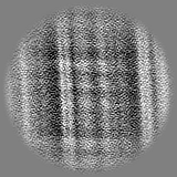
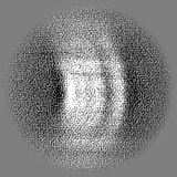
| Projections & Slices |
| ||||||||||||
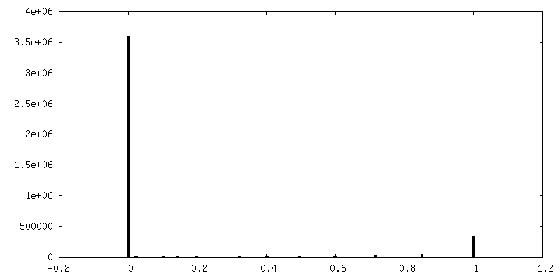
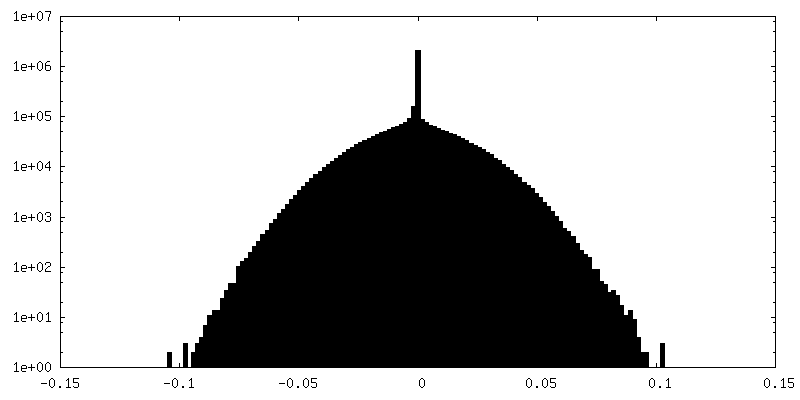
| Density Histograms |
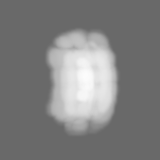

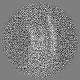
-Half map: half map 1 of sub-tomogram average of 4-beta-ring hoop
| File | emd_29442_half_map_1.map | ||||||||||||
|---|---|---|---|---|---|---|---|---|---|---|---|---|---|
| Annotation | half map 1 of sub-tomogram average of 4-beta-ring hoop | ||||||||||||
| Projections & Slices |
| ||||||||||||
| Density Histograms |

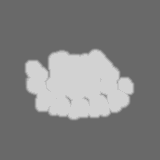
-Half map: half map 2 of sub-tomogram average of 4-beta-ring hoop
| File | emd_29442_half_map_2.map | ||||||||||||
|---|---|---|---|---|---|---|---|---|---|---|---|---|---|
| Annotation | half map 2 of sub-tomogram average of 4-beta-ring hoop | ||||||||||||
| Projections & Slices |
| ||||||||||||
| Density Histograms |


- Sample components
-Entire : Methanospirillum hungatei
| Entire | Name: Methanospirillum hungatei |
|---|---|
| Components |
|
-Supramolecule #1: Methanospirillum hungatei
| Supramolecule | Name: Methanospirillum hungatei / type: cell / ID: 1 / Parent: 0 / Details: the whole cell of Methanospirillum hungatei |
|---|---|
| Source (natural) | Organism: Methanospirillum hungatei JF-1 (archaea) |
-Experimental details
-Structure determination
| Method | cryo EM |
|---|---|
 Processing Processing | subtomogram averaging |
| Aggregation state | cell |
-Sample preparation
| Buffer | pH: 7 |
|---|---|
| Vitrification | Cryogen name: ETHANE |
- Electron microscopy
| Microscope | FEI TITAN KRIOS |
|---|---|
| Image recording | Film or detector model: GATAN K2 SUMMIT (4k x 4k) / Detector mode: COUNTING / Average electron dose: 1.8 e/Å2 |
| Electron beam | Acceleration voltage: 300 kV / Electron source:  FIELD EMISSION GUN FIELD EMISSION GUN |
| Electron optics | Illumination mode: FLOOD BEAM / Imaging mode: BRIGHT FIELD / Nominal defocus max: 4.0 µm / Nominal defocus min: 1.5 µm |
| Experimental equipment |  Model: Titan Krios / Image courtesy: FEI Company |
-Image processing
| Final reconstruction | Applied symmetry - Point group: C1 (asymmetric) / Resolution.type: BY AUTHOR / Resolution: 7.92 Å / Resolution method: FSC 0.143 CUT-OFF / Software - Name: RELION (ver. 4.0) Details: The FSC curve was performed on 3dfsc server https://3dfsc.salk.edu/ Number subtomograms used: 17359 |
|---|---|
| Extraction | Number tomograms: 10 / Number images used: 20137 |
| Final angle assignment | Type: ANGULAR RECONSTITUTION |
