Movie
Movie Controller

[English] 日本語
 Yorodumi
- EMDB-29027: CryoEM structure of bacteriophage Q-beta coat protein dimer with ... -
+ Open data

- Basic information
| Entry |  | |||||||||
|---|---|---|---|---|---|---|---|---|---|---|
| Title | CryoEM structure of bacteriophage Q-beta coat protein dimer with AYGG linker | |||||||||
 Map data Map data | Final map | |||||||||
 Sample Sample |
| |||||||||
 Keywords Keywords | coat protein / q-beta / fusion / VIRUS LIKE PARTICLE | |||||||||
| Function / homology | Read-through domain / Read-through domain / Levivirus coat protein / Levivirus coat protein / Bacteriophage RNA-type, capsid / viral capsid / structural molecule activity / Minor capsid protein A1 Function and homology information Function and homology information | |||||||||
| Biological species |  Qubevirus durum Qubevirus durum | |||||||||
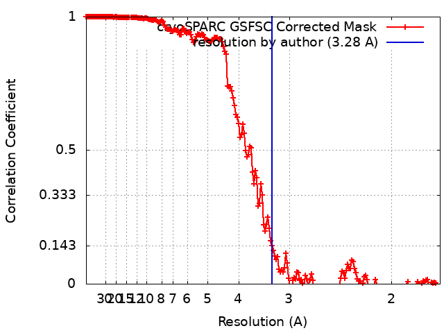
| Method | single particle reconstruction / cryo EM / Resolution: 3.28 Å | |||||||||
 Authors Authors | Newton TP / Zhao L / Finn MG / Kopylov M | |||||||||
| Funding support |  United States, 1 items United States, 1 items
| |||||||||
 Citation Citation | Journal: To Be Published Title: CryoEM structure of bacteriophage Q-beta coat protein dimer with AYGG linker Authors: Newton TP / Zhao L / Kopylov M / Finn MG | |||||||||
| History |
|
- Structure visualization
| Supplemental images |
|---|

- Downloads & links
-EMDB archive
| Map data | emd_29027.map.gz | 255 MB | EMDB map data format | |
|---|---|---|---|---|
| Header (meta data) | emd-29027-v30.xmlemd-29027.xml | 13.8 KB 13.8 KB | Display Display | EMDB header |
| FSC (resolution estimation) | emd_29027_fsc.xml | 16.9 KB | Display | FSC data file |
| Images | 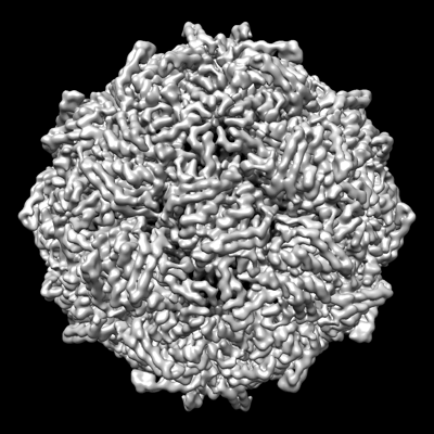 emd_29027.png emd_29027.png | 142.6 KB | ||
| Masks | emd_29027_msk_1.map | 512 MB | Mask map | |
| Filedesc metadata | emd-29027.cif.gz | 5.2 KB | ||
| Others | emd_29027_half_map_1.map.gzemd_29027_half_map_2.map.gz | 475.6 MB 475.5 MB | ||
| Archive directory |  http://ftp.pdbj.org/pub/emdb/structures/EMD-29027ftp://ftp.pdbj.org/pub/emdb/structures/EMD-29027 http://ftp.pdbj.org/pub/emdb/structures/EMD-29027ftp://ftp.pdbj.org/pub/emdb/structures/EMD-29027 | HTTPS FTP |
-Related structure data
| Related structure data | 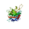 8fehMC M: atomic model generated by this map C: citing same article ( |
|---|---|
| Similar structure data |
-Links
| EMDB pages | EMDB (EBI/PDBe) / EMDataResource |
|---|
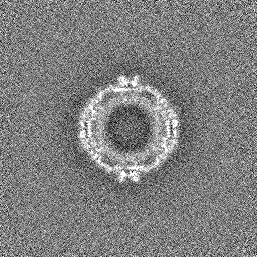
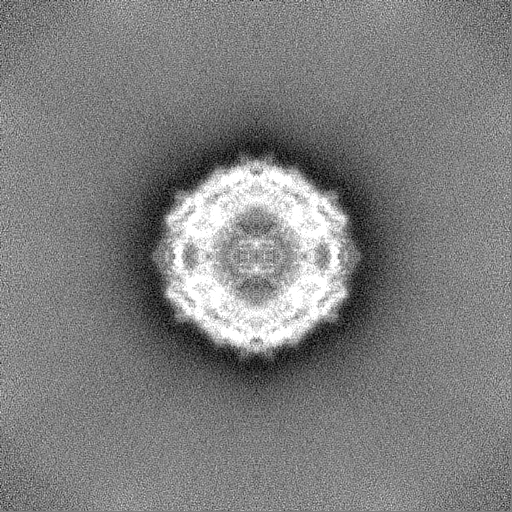
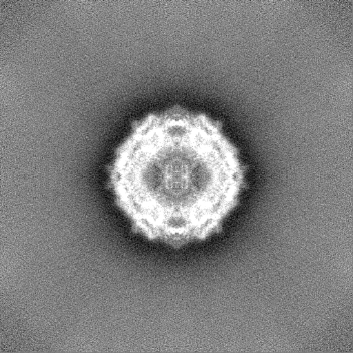
-Map
| File | Download / File: emd_29027.map.gz / Format: CCP4 / Size: 512 MB / Type: IMAGE STORED AS FLOATING POINT NUMBER (4 BYTES) | ||||||||||||||||||||||||||||||||||||
|---|---|---|---|---|---|---|---|---|---|---|---|---|---|---|---|---|---|---|---|---|---|---|---|---|---|---|---|---|---|---|---|---|---|---|---|---|---|
| Annotation | Final map | ||||||||||||||||||||||||||||||||||||
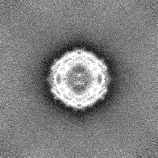
| Projections & slices | Image control
Images are generated by Spider. | ||||||||||||||||||||||||||||||||||||
| Voxel size | X=Y=Z: 0.855 Å | ||||||||||||||||||||||||||||||||||||
| Density |
| ||||||||||||||||||||||||||||||||||||
| Symmetry | Space group: 1 | ||||||||||||||||||||||||||||||||||||
| Details | EMDB XML:
|
 Z (Sec.)
 Y (Row.)
 X (Col.)


-Supplemental data
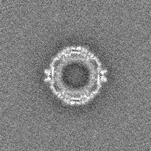
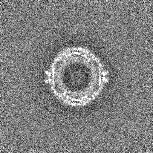
-Mask #1
| File | emd_29027_msk_1.map | ||||||||||||
|---|---|---|---|---|---|---|---|---|---|---|---|---|---|
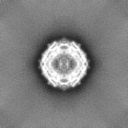
| Projections & Slices |
| ||||||||||||
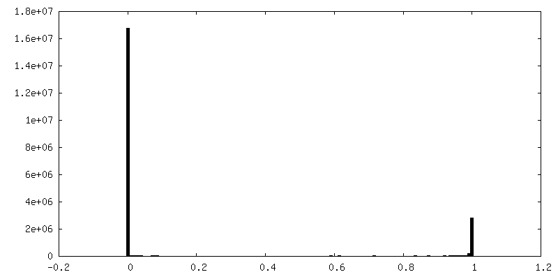
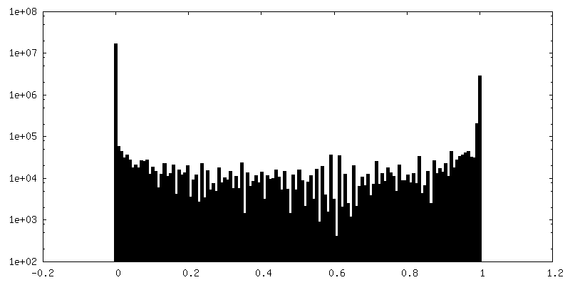
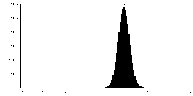
| Density Histograms |


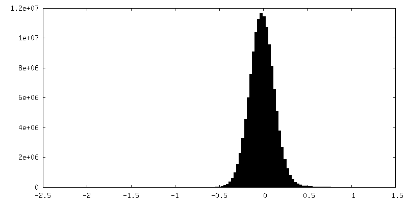
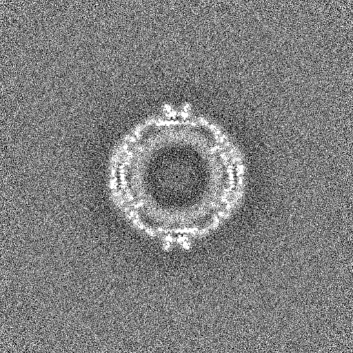
-Half map: Final half map A
| File | emd_29027_half_map_1.map | ||||||||||||
|---|---|---|---|---|---|---|---|---|---|---|---|---|---|
| Annotation | Final half map A | ||||||||||||
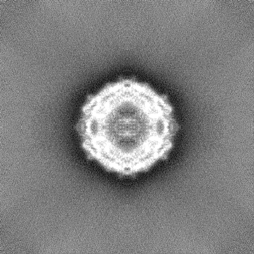
| Projections & Slices |
| ||||||||||||
| Density Histograms |

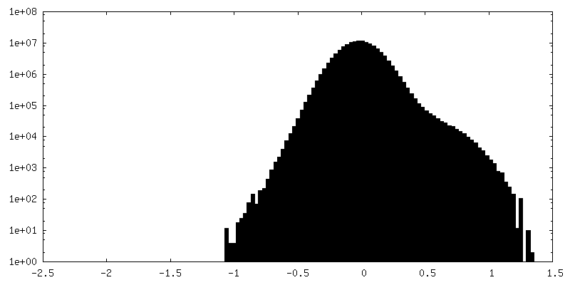
-Half map: Final half map B
| File | emd_29027_half_map_2.map | ||||||||||||
|---|---|---|---|---|---|---|---|---|---|---|---|---|---|
| Annotation | Final half map B | ||||||||||||
| Projections & Slices |
| ||||||||||||
| Density Histograms |


- Sample components
-Entire : Qubevirus durum
| Entire | Name: Qubevirus durum |
|---|---|
| Components |
|
-Supramolecule #1: Qubevirus durum
| Supramolecule | Name: Qubevirus durum / type: virus / ID: 1 / Parent: 0 / Macromolecule list: all / NCBI-ID: 39803 / Sci species name: Qubevirus durum / Virus type: VIRUS-LIKE PARTICLE / Virus isolate: OTHER / Virus enveloped: No / Virus empty: Yes |
|---|
-Macromolecule #1: Minor capsid protein A1 fusion
| Macromolecule | Name: Minor capsid protein A1 fusion / type: protein_or_peptide / ID: 1 / Number of copies: 1 / Enantiomer: LEVO |
|---|---|
| Source (natural) | Organism: Qubevirus durum |
| Molecular weight | Theoretical: 28.604137 KDa |
| Recombinant expression | Organism:  |
| Sequence | String: AKLETVTLGN IGKDGKQTLV LNPRGVNPTN GVASLSQAGA VPALEKRVTV SVSQPSRNRK NYKVQVKIQN PTACTANGSC DPSVTRQAY ADVTFSFTQY STDEERAFVR TELAALLASP LLIDAIDQLN PAYAYGGAKL ETVTLGNIGK DGKQTLVLNP R GVNPTNGV ...String: AKLETVTLGN IGKDGKQTLV LNPRGVNPTN GVASLSQAGA VPALEKRVTV SVSQPSRNRK NYKVQVKIQN PTACTANGSC DPSVTRQAY ADVTFSFTQY STDEERAFVR TELAALLASP LLIDAIDQLN PAYAYGGAKL ETVTLGNIGK DGKQTLVLNP R GVNPTNGV ASLSQAGAVP ALEKRVTVSV SQPSRNRKNY KVQVKIQNPT ACTANGSCDP SVTRQAYADV TFSFTQYSTD EE RAFVRTE LAALLASPLL IDAIDQLNPA Y UniProtKB: Minor capsid protein A1 |
-Experimental details
-Structure determination
| Method | cryo EM |
|---|---|
 Processing Processing | single particle reconstruction |
| Aggregation state | particle |
-Sample preparation
| Concentration | 1 mg/mL |
|---|---|
| Buffer | pH: 7.5 |
| Vitrification | Cryogen name: ETHANE |
- Electron microscopy
| Microscope | TFS KRIOS |
|---|---|
| Image recording | Film or detector model: GATAN K2 SUMMIT (4k x 4k) / Detector mode: COUNTING / Digitization - Dimensions - Width: 3838 pixel / Digitization - Dimensions - Height: 3710 pixel / Average electron dose: 64.7 e/Å2 |
| Electron beam | Acceleration voltage: 300 kV / Electron source:  FIELD EMISSION GUN FIELD EMISSION GUN |
| Electron optics | C2 aperture diameter: 100.0 µm / Illumination mode: FLOOD BEAM / Imaging mode: BRIGHT FIELD / Cs: 0.001 mm / Nominal defocus max: 2.0 µm / Nominal defocus min: 1.0 µm |
| Experimental equipment |  Model: Titan Krios / Image courtesy: FEI Company |