 Movie
Movie Controller

[English] 日本語
 Yorodumi
- EMDB-28906: Thermoplasma acidophilum 20S proteasome - wild type bound to ZYA -
+ Open data

- Basic information
| Entry |  | |||||||||
|---|---|---|---|---|---|---|---|---|---|---|
| Title | Thermoplasma acidophilum 20S proteasome - wild type bound to ZYA | |||||||||
 Map data Map data | T20S ZYA EM Map unsharpened | |||||||||
 Sample Sample |
| |||||||||
 Keywords Keywords | Protease / threonine protease / endopeptidase activity / HYDROLASE | |||||||||
| Function / homology |  Function and homology information Function and homology informationproteasome endopeptidase complex / proteasome core complex, beta-subunit complex / threonine-type endopeptidase activity / proteasome core complex, alpha-subunit complex / proteasomal protein catabolic process / endopeptidase activity / ubiquitin-dependent protein catabolic process / cytoplasm Similarity search - Function | |||||||||
| Biological species |   Thermoplasma acidophilum (acidophilic) Thermoplasma acidophilum (acidophilic) | |||||||||
| Method | single particle reconstruction / cryo EM / Resolution: 1.94 Å | |||||||||
 Authors Authors | Chuah J / Smith D | |||||||||
| Funding support |  United States, 1 items United States, 1 items
| |||||||||
 Citation Citation | Journal: Commun Biol / Year: 2023 Title: High resolution structures define divergent and convergent mechanisms of archaeal proteasome activation. Authors: Janelle J Y Chuah / Matthew S Rexroad / David M Smith / Abstract: Considering the link between neurodegenerative diseases and impaired proteasome function, and the neuro-protective impact of enhanced proteasome activity in animal models, it's crucial to understand ...Considering the link between neurodegenerative diseases and impaired proteasome function, and the neuro-protective impact of enhanced proteasome activity in animal models, it's crucial to understand proteasome activation mechanisms. A hydrophobic-tyrosine-any residue (HbYX) motif on the C-termini of proteasome-activating complexes independently triggers gate-opening of the 20S core particle for protein degradation; however, the causal allosteric mechanism remains unclear. Our study employs a structurally irreducible dipeptide HbYX mimetic to investigate the allosteric mechanism of gate-opening in the archaeal proteasome. High-resolution cryo-EM structures pinpoint vital residues and conformational changes in the proteasome α-subunit implicated in HbYX-dependent activation. Using point mutations, we simulated the HbYX-bound state, providing support for our mechanistic model. We discerned four main mechanistic elements triggering gate-opening: 1) back-loop rearrangement adjacent to K66, 2) intra- and inter- α subunit conformational changes, 3) occupancy of the hydrophobic pocket, and 4) a highly conserved isoleucine-threonine pair in the 20S channel stabilizing the open and closed states, termed the "IT switch." Comparison of different complexes unveiled convergent and divergent mechanism of 20S gate-opening among HbYX-dependent and independent activators. This study delivers a detailed molecular model for HbYX-dependent 20S gate-opening, enabling the development of small molecule proteasome activators that hold promise to treat neurodegenerative diseases. | |||||||||
| History |
|
- Structure visualization
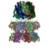
| Supplemental images |
|---|

- Downloads & links
-EMDB archive
| Map data | emd_28906.map.gz | 163.3 MB | EMDB map data format | |
|---|---|---|---|---|
| Header (meta data) | emd-28906-v30.xmlemd-28906.xml | 17.5 KB 17.5 KB | Display Display | EMDB header |
| FSC (resolution estimation) | emd_28906_fsc.xml | 14.4 KB | Display | FSC data file |
| Images | 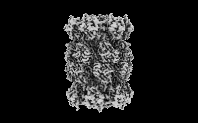 emd_28906.png emd_28906.png | 49.7 KB | ||
| Others | emd_28906_additional_1.map.gzemd_28906_half_map_1.map.gzemd_28906_half_map_2.map.gz | 33 MB 301.7 MB 301.7 MB | ||
| Archive directory |  http://ftp.pdbj.org/pub/emdb/structures/EMD-28906ftp://ftp.pdbj.org/pub/emdb/structures/EMD-28906 http://ftp.pdbj.org/pub/emdb/structures/EMD-28906ftp://ftp.pdbj.org/pub/emdb/structures/EMD-28906 | HTTPS FTP |
-Related structure data
| Related structure data |  8f7kMC  8f66C  8f6aC M: atomic model generated by this map C: citing same article ( |
|---|---|
| Similar structure data |


-Links
| EMDB pages | EMDB (EBI/PDBe) / EMDataResource |
|---|---|
| Related items in Molecule of the Month |


-Map
| File | Download / File: emd_28906.map.gz / Format: CCP4 / Size: 325 MB / Type: IMAGE STORED AS FLOATING POINT NUMBER (4 BYTES) | ||||||||||||||||||||||||||||||||||||
|---|---|---|---|---|---|---|---|---|---|---|---|---|---|---|---|---|---|---|---|---|---|---|---|---|---|---|---|---|---|---|---|---|---|---|---|---|---|
| Annotation | T20S ZYA EM Map unsharpened | ||||||||||||||||||||||||||||||||||||
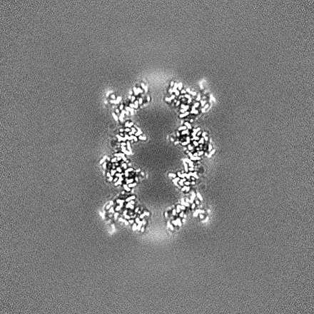
| Projections & slices | Image control
Images are generated by Spider. | ||||||||||||||||||||||||||||||||||||
| Voxel size | X=Y=Z: 0.73636 Å | ||||||||||||||||||||||||||||||||||||
| Density |
| ||||||||||||||||||||||||||||||||||||
| Symmetry | Space group: 1 | ||||||||||||||||||||||||||||||||||||
| Details | EMDB XML:
|
 Z (Sec.)
 Y (Row.)
 X (Col.)


-Supplemental data
-Additional map: T20S ZYA Denmod Map
| File | emd_28906_additional_1.map | ||||||||||||
|---|---|---|---|---|---|---|---|---|---|---|---|---|---|
| Annotation | T20S ZYA Denmod Map | ||||||||||||
| Projections & Slices |
| ||||||||||||
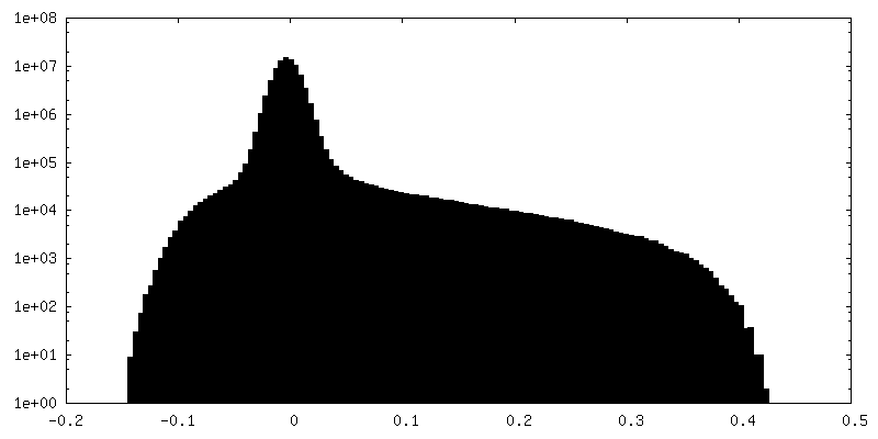
| Density Histograms |


-Half map: T20S ZYA EM Map Half A
| File | emd_28906_half_map_1.map | ||||||||||||
|---|---|---|---|---|---|---|---|---|---|---|---|---|---|
| Annotation | T20S ZYA EM Map Half A | ||||||||||||
| Projections & Slices |
| ||||||||||||
| Density Histograms |


-Half map: T20S ZYA EM Map Half B
| File | emd_28906_half_map_2.map | ||||||||||||
|---|---|---|---|---|---|---|---|---|---|---|---|---|---|
| Annotation | T20S ZYA EM Map Half B | ||||||||||||
| Projections & Slices |
| ||||||||||||
| Density Histograms |


- Sample components
-Entire : Wild-type Thermoplasma acidophilum 20S proteasome bound to ZYA
| Entire | Name: Wild-type Thermoplasma acidophilum 20S proteasome bound to ZYA |
|---|---|
| Components |
|
-Supramolecule #1: Wild-type Thermoplasma acidophilum 20S proteasome bound to ZYA
| Supramolecule | Name: Wild-type Thermoplasma acidophilum 20S proteasome bound to ZYA type: complex / ID: 1 / Parent: 0 / Macromolecule list: #2, #1 |
|---|---|
| Source (natural) | Organism: Thermoplasma acidophilum (acidophilic) |
| Molecular weight | Theoretical: 700 kDa/nm |
-Macromolecule #1: Proteasome subunit alpha
| Macromolecule | Name: Proteasome subunit alpha / type: protein_or_peptide / ID: 1 / Number of copies: 14 / Enantiomer: LEVO |
|---|---|
| Source (natural) | Organism: Thermoplasma acidophilum (acidophilic) |
| Molecular weight | Theoretical: 25.441996 KDa |
| Recombinant expression | Organism:  |
| Sequence | String: GQMAYDRAIT VFSPDGRLFQ VEYAREAVKK GSTALGMKFA NGVLLISDKK VRSRLIEQNS IEKIQLIDDY VAAVTSGLVA DARVLVDFA RISAQQEKVT YGSLVNIENL VKRVADQMQQ YTQYGGVRPY GVSLIFAGID QIGPRLFDCD PAGTINEYKA T AIGSGKDA ...String: GQMAYDRAIT VFSPDGRLFQ VEYAREAVKK GSTALGMKFA NGVLLISDKK VRSRLIEQNS IEKIQLIDDY VAAVTSGLVA DARVLVDFA RISAQQEKVT YGSLVNIENL VKRVADQMQQ YTQYGGVRPY GVSLIFAGID QIGPRLFDCD PAGTINEYKA T AIGSGKDA VVSFLEREYK ENLPEKEAVT LGIKALKSSL EEGEELKAPE IASITVGNKY RIYDQEEVKK FL UniProtKB: Proteasome subunit alpha |
-Macromolecule #2: Proteasome subunit beta
| Macromolecule | Name: Proteasome subunit beta / type: protein_or_peptide / ID: 2 / Number of copies: 14 / Enantiomer: LEVO / EC number: proteasome endopeptidase complex |
|---|---|
| Source (natural) | Organism: Thermoplasma acidophilum (acidophilic) |
| Molecular weight | Theoretical: 22.294848 KDa |
| Recombinant expression | Organism: |
| Sequence | String: TTTVGITLKD AVIMATERRV TMENFIMHKN GKKLFQIDTY TGMTIAGLVG DAQVLVRYMK AELELYRLQR RVNMPIEAVA TLLSNMLNQ VKYMPYMVQL LVGGIDTAPH VFSIDAAGGS VEDIYASTGS GSPFVYGVLE SQYSEKMTVD EGVDLVIRAI S AAKQRDSA ...String: TTTVGITLKD AVIMATERRV TMENFIMHKN GKKLFQIDTY TGMTIAGLVG DAQVLVRYMK AELELYRLQR RVNMPIEAVA TLLSNMLNQ VKYMPYMVQL LVGGIDTAPH VFSIDAAGGS VEDIYASTGS GSPFVYGVLE SQYSEKMTVD EGVDLVIRAI S AAKQRDSA SGGMIDVAVI TRKDGYVQLP TDQIESRIRK LGLIL UniProtKB: Proteasome subunit beta |
-Macromolecule #3: N-[(benzyloxy)carbonyl]-L-tyrosyl-D-alanine
| Macromolecule | Name: N-[(benzyloxy)carbonyl]-L-tyrosyl-D-alanine / type: ligand / ID: 3 / Number of copies: 14 / Formula: XIB |
|---|---|
| Molecular weight | Theoretical: 386.398 Da |
| Chemical component information |  ChemComp-XIB: |
-Experimental details
-Structure determination
| Method | cryo EM |
|---|---|
 Processing Processing | single particle reconstruction |
| Aggregation state | particle |
-Sample preparation
| Concentration | 1 mg/mL |
|---|---|
| Buffer | pH: 7.4 |
| Vitrification | Cryogen name: ETHANE |
- Electron microscopy
| Microscope | TFS KRIOS |
|---|---|
| Image recording | Film or detector model: FEI FALCON III (4k x 4k) / Detector mode: SUPER-RESOLUTION / Average electron dose: 50.0 e/Å2 |
| Electron beam | Acceleration voltage: 300 kV / Electron source:  FIELD EMISSION GUN FIELD EMISSION GUN |
| Electron optics | Illumination mode: FLOOD BEAM / Imaging mode: BRIGHT FIELD / Nominal defocus max: 2.4 µm / Nominal defocus min: 1.2 µm |
| Experimental equipment |  Model: Titan Krios / Image courtesy: FEI Company |
+Image processing

-Atomic model buiding 1
| Refinement | Protocol: FLEXIBLE FIT |
|---|---|
| Output model | PDB-8f7k: |
