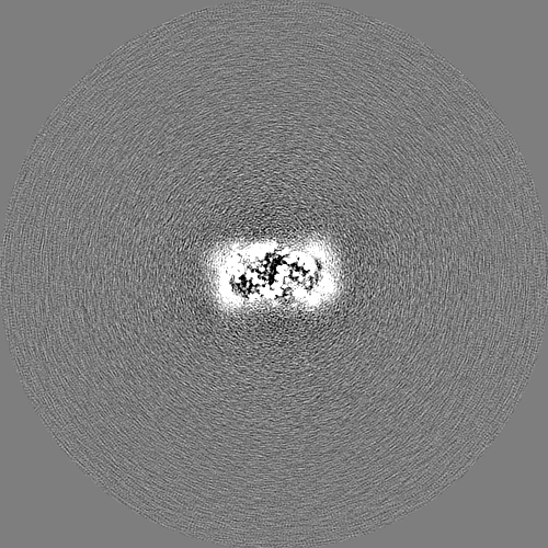
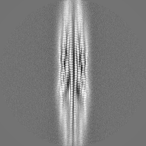
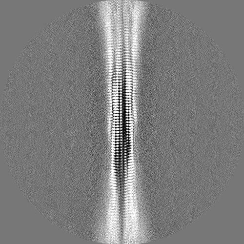
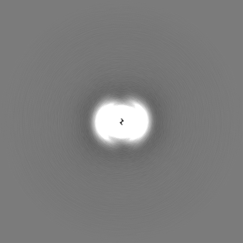
Journal: Proc Natl Acad Sci U S A / Year: 2023 Title: Structures of brain-derived 42-residue amyloid-β fibril polymorphs with unusual molecular conformations and intermolecular interactions. Authors: Myungwoon Lee / Wai-Ming Yau / John M Louis / Robert Tycko / Abstract: Fibrils formed by the 42-residue amyloid-β peptide (Aβ42), a main component of amyloid deposits in Alzheimer's disease (AD), are known to be polymorphic, i.e., to contain multiple possible ...Fibrils formed by the 42-residue amyloid-β peptide (Aβ42), a main component of amyloid deposits in Alzheimer's disease (AD), are known to be polymorphic, i.e., to contain multiple possible molecular structures. Previous studies of Aβ42 fibrils, including fibrils prepared entirely in vitro or extracted from brain tissue and using solid-state NMR (ssNMR) or cryogenic electron microscopy (cryo-EM) methods, have found polymorphs with differences in amino acid sidechain orientations, lengths of structurally ordered segments, and contacts between cross-β subunit pairs within a single filament. Despite these differences, Aβ42 molecules adopt a common S-shaped conformation in all previously described high-resolution Aβ42 fibril structures. Here we report two cryo-EM-based structures of Aβ42 fibrils that are qualitatively different, in samples derived from AD brain tissue by seeded growth. In type A fibrils, residues 12 to 42 adopt a ν-shaped conformation, with both intra-subunit and intersubunit hydrophobic contacts to form a compact core. In type B fibrils, residues 2 to 42 adopt an υ-shaped conformation, with only intersubunit contacts and internal pores. Type A and type B fibrils have opposite helical handedness. Cryo-EM density maps and molecular dynamics simulations indicate intersubunit K16-A42 salt bridges in type B fibrils and partially occupied K28-A42 salt bridges in type A fibrils. The coexistence of two predominant polymorphs, with differences in N-terminal dynamics, is supported by ssNMR data, as is faithful propagation of structures from first-generation to second-generation brain-seeded Aβ42 fibril samples. These results demonstrate that Aβ42 fibrils can exhibit a greater range of structural variations than seen in previous studies.
Model: Quantifoil R1.2/1.3 / Material: COPPER / Mesh: 400 / Pretreatment - Type: GLOW DISCHARGE / Pretreatment - Time: 45 sec. / Pretreatment - Pressure: 0.039 kPa Details: The grid was glow discharged immediately before use.
Vitrification
Cryogen name: ETHANE / Chamber humidity: 99 % / Chamber temperature: 93 K / Instrument: FEI VITROBOT MARK I Details: Preblot for 12-13 seconds and blot for 2.5-3.0 seconds before plunging.
-
Electron microscopy
Microscope
FEI TITAN KRIOS
Image recording
Film or detector model: GATAN K2 SUMMIT (4k x 4k) / Detector mode: SUPER-RESOLUTION / Digitization - Dimensions - Width: 11520 pixel / Digitization - Dimensions - Height: 8184 pixel / Number grids imaged: 1 / Number real images: 5557 / Average exposure time: 1.65 sec. / Average electron dose: 50.34 e/Å2
Electron beam
Acceleration voltage: 300 kV / Electron source: FIELD EMISSION GUN
In the structure databanks used in Yorodumi, some data are registered as the other names, "COVID-19 virus" and "2019-nCoV". Here are the details of the virus and the list of structure data.
Jan 31, 2019. EMDB accession codes are about to change! (news from PDBe EMDB page)
EMDB accession codes are about to change! (news from PDBe EMDB page)
The allocation of 4 digits for EMDB accession codes will soon come to an end. Whilst these codes will remain in use, new EMDB accession codes will include an additional digit and will expand incrementally as the available range of codes is exhausted. The current 4-digit format prefixed with “EMD-” (i.e. EMD-XXXX) will advance to a 5-digit format (i.e. EMD-XXXXX), and so on. It is currently estimated that the 4-digit codes will be depleted around Spring 2019, at which point the 5-digit format will come into force.
The EM Navigator/Yorodumi systems omit the EMD- prefix.
Related info.:Q: What is EMD? / ID/Accession-code notation in Yorodumi/EM Navigator
Yorodumi is a browser for structure data from EMDB, PDB, SASBDB, etc.
This page is also the successor to EM Navigator detail page, and also detail information page/front-end page for Omokage search.
The word "yorodu" (or yorozu) is an old Japanese word meaning "ten thousand". "mi" (miru) is to see.
Related info.:EMDB / PDB / SASBDB / Comparison of 3 databanks / Yorodumi Search / Aug 31, 2016. New EM Navigator & Yorodumi / Yorodumi Papers / Jmol/JSmol / Function and homology information / Changes in new EM Navigator and Yorodumi
 Movie
Movie Controller


 Open data
 Basic information
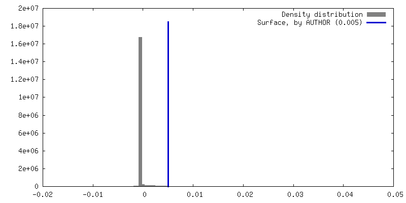
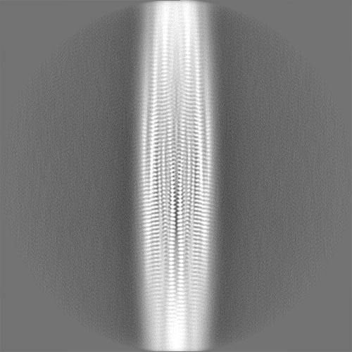
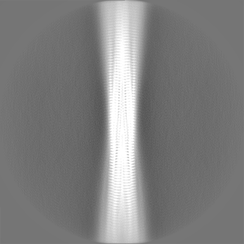
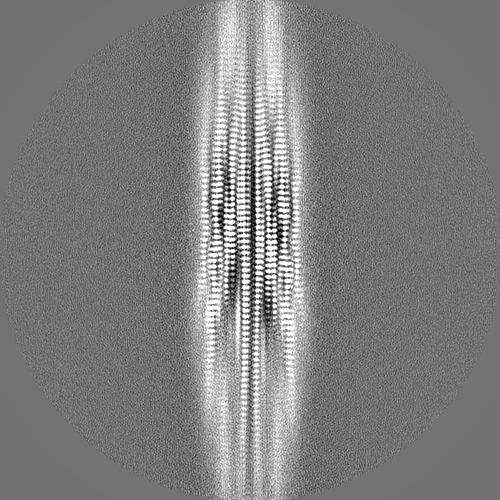
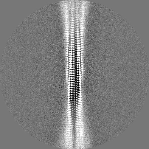
 Map data
 Sample
 Keywords
 Function and homology information
 Homo sapiens (human)
 Authors
 United States, 1 items
  Citation
 Structure visualization
 Downloads & links
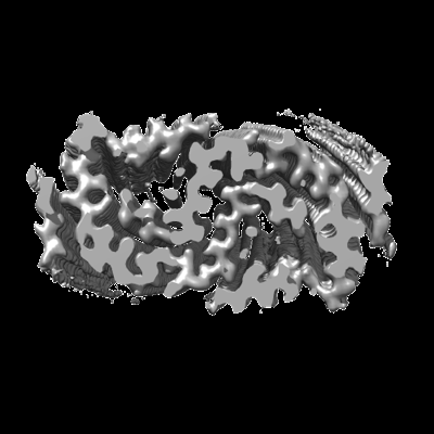 emd_28741.png
 http://ftp.pdbj.org/pub/emdb/structures/EMD-28741


 Z (Sec.)
 Y (Row.)
 X (Col.)


 Sample components
 Processing
 Electron microscopy
 FIELD EMISSION GUN
