[English] 日本語
Yorodumi
- EMDB-28554: CryoEM Structure of Lipoprotein Lipase Dimer -

+
Open data


ID or keywords:

Loading...

-
Basic information

Entry
Database: EMDB / ID: EMD-28554
TitleCryoEM Structure of Lipoprotein Lipase Dimer
Map data
Sample
  • Complex: Lipoprotein Lipase Dimer
    • Protein or peptide: Lipoprotein lipase
KeywordsDimer / lipase / HYDROLASE
Function / homology
Function and homology information


Assembly of active LPL and LIPC lipase complexes / positive regulation of chemokine production => GO:0032722 / lipoprotein lipase / lipoprotein lipase activity / positive regulation of tumor necrosis factor production => GO:0032760 / positive regulation of sequestering of triglyceride / low-density lipoprotein particle mediated signaling / chylomicron remodeling / positive regulation of cholesterol storage / phospholipase A1 ...Assembly of active LPL and LIPC lipase complexes / positive regulation of chemokine production => GO:0032722 / lipoprotein lipase / lipoprotein lipase activity / positive regulation of tumor necrosis factor production => GO:0032760 / positive regulation of sequestering of triglyceride / low-density lipoprotein particle mediated signaling / chylomicron remodeling / positive regulation of cholesterol storage / phospholipase A1 / phosphatidylserine 1-acylhydrolase activity / 1-acyl-2-lysophosphatidylserine acylhydrolase activity / phospholipase A1 activity / triglyceride catabolic process / phospholipase activity / lipase activity / very-low-density lipoprotein particle remodeling / positive regulation of macrophage derived foam cell differentiation / cellular response to nutrient / chylomicron / very-low-density lipoprotein particle / triacylglycerol lipase activity / heparan sulfate proteoglycan binding / positive regulation of chemokine (C-X-C motif) ligand 2 production / triglyceride homeostasis / cellular response to fatty acid / triglyceride metabolic process / lipoprotein particle binding / apolipoprotein binding / positive regulation of fat cell differentiation / phospholipid metabolic process / response to glucose / lipid catabolic process / positive regulation of interleukin-1 beta production / cholesterol homeostasis / response to bacterium / fatty acid biosynthetic process / positive regulation of inflammatory response / positive regulation of interleukin-6 production / heparin binding / signaling receptor binding / calcium ion binding / cell surface / protein homodimerization activity / extracellular space / plasma membrane
Similarity search - Function
Lipoprotein lipase / Lipase, LIPH-type / Lipase, N-terminal / Triacylglycerol lipase family / Lipase / Lipase / Lipoxygenase homology 2 (beta barrel) domain / PLAT/LH2 domain / PLAT/LH2 domain superfamily / PLAT/LH2 domain ...Lipoprotein lipase / Lipase, LIPH-type / Lipase, N-terminal / Triacylglycerol lipase family / Lipase / Lipase / Lipoxygenase homology 2 (beta barrel) domain / PLAT/LH2 domain / PLAT/LH2 domain superfamily / PLAT/LH2 domain / PLAT domain profile. / Lipases, serine active site. / Alpha/Beta hydrolase fold
Similarity search - Domain/homology
Biological speciesBos taurus (cattle) / cattle (cattle)
Methodsingle particle reconstruction / cryo EM / Resolution: 3.9 Å
AuthorsGunn KH / Neher SB
Funding support United States, 2 items
OrganizationGrant numberCountry
National Institutes of Health/National Heart, Lung, and Blood Institute (NIH/NHLBI)R01HL125654 United States
American Heart Association900354 United States
Citation
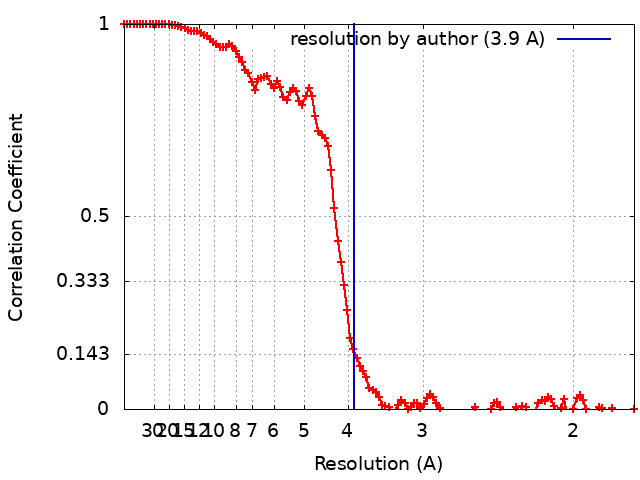
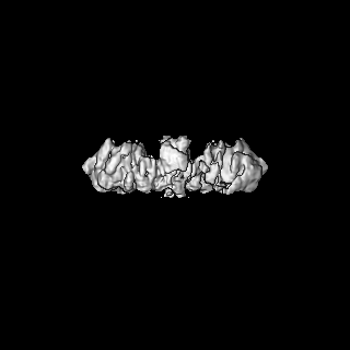
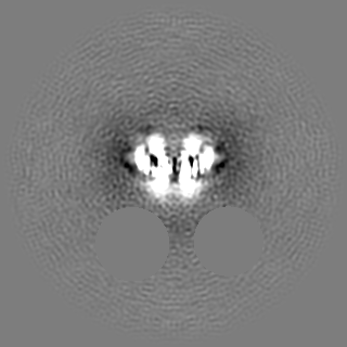
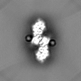
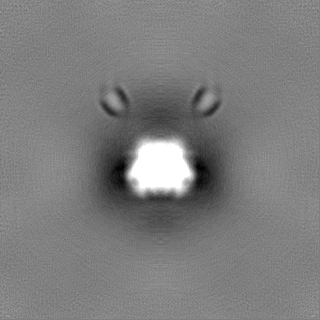
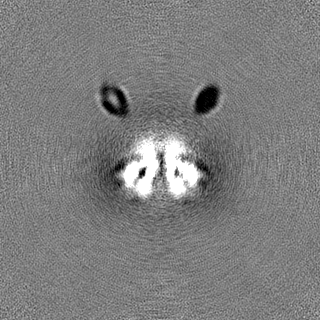
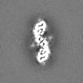
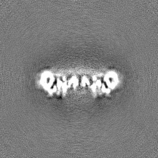
Journal: Nat Commun / Year: 2023
Title: Structure of dimeric lipoprotein lipase reveals a pore adjacent to the active site.
Authors: Kathryn H Gunn / Saskia B Neher /
Abstract: Lipoprotein lipase (LPL) hydrolyzes triglycerides from circulating lipoproteins, releasing free fatty acids. Active LPL is needed to prevent hypertriglyceridemia, which is a risk factor for ...Lipoprotein lipase (LPL) hydrolyzes triglycerides from circulating lipoproteins, releasing free fatty acids. Active LPL is needed to prevent hypertriglyceridemia, which is a risk factor for cardiovascular disease (CVD). Using cryogenic electron microscopy (cryoEM), we determined the structure of an active LPL dimer at 3.9 Å resolution. This structure reveals an open hydrophobic pore adjacent to the active site residues. Using modeling, we demonstrate that this pore can accommodate an acyl chain from a triglyceride. Known LPL mutations that lead to hypertriglyceridemia localize to the end of the pore and cause defective substrate hydrolysis. The pore may provide additional substrate specificity and/or allow unidirectional acyl chain release from LPL. This structure also revises previous models on how LPL dimerizes, revealing a C-terminal to C-terminal interface. We hypothesize that this active C-terminal to C-terminal conformation is adopted by LPL when associated with lipoproteins in capillaries.
History
DepositionOct 12, 2022-
Header (metadata) releaseMay 3, 2023-
Map releaseMay 3, 2023-
UpdateJun 14, 2023-
Current statusJun 14, 2023Processing site: RCSB / Status: Released

-
Structure visualization

Supplemental images

Downloads & links

-
Map

FileDownload / File: emd_28554.map.gz / Format: CCP4 / Size: 125 MB / Type: IMAGE STORED AS FLOATING POINT NUMBER (4 BYTES)
Voxel sizeX=Y=Z: 0.88 Å
Density
Contour LevelBy AUTHOR: 0.05
Minimum - Maximum-0.17224558 - 0.37482417
Average (Standard dev.)-0.00017229572 (±0.0082041295)
SymmetrySpace group: 1
Details

EMDB XML:

Map geometry
Axis orderXYZ
Origin000
Dimensions320320320
Spacing320320320
CellA=B=C: 281.6 Å
α=β=γ: 90.0 °

-
Supplemental data

-
Half map: #2

Fileemd_28554_half_map_1.map
Projections & Slices
AxesZYX

Projections

Slices (1/2)
Density Histograms

-
Half map: #1

Fileemd_28554_half_map_2.map
Projections & Slices
AxesZYX

Projections

Slices (1/2)
Density Histograms

-
Sample components

-
Entire : Lipoprotein Lipase Dimer

EntireName: Lipoprotein Lipase Dimer
Components
  • Complex: Lipoprotein Lipase Dimer
    • Protein or peptide: Lipoprotein lipase

-
Supramolecule #1: Lipoprotein Lipase Dimer

SupramoleculeName: Lipoprotein Lipase Dimer / type: complex / ID: 1 / Parent: 0 / Macromolecule list: all
Source (natural)Organism: Bos taurus (cattle)
Molecular weightTheoretical: 101 KDa

-
Macromolecule #1: Lipoprotein lipase

MacromoleculeName: Lipoprotein lipase / type: protein_or_peptide / ID: 1 / Number of copies: 2 / Enantiomer: LEVO / EC number: lipoprotein lipase
Source (natural)Organism: cattle (cattle)
Molecular weightTheoretical: 53.448789 KDa
SequenceString: MESKALLLLA LSVCLQSLTV SRGGLVAADR ITGGKDFRDI ESKFALRTPE DTAEDTCHLI PGVTESVANC HFNHSSKTFV VIHGWTVTG MYESWVPKLV AALYKREPDS NVIVVDWLSR AQQHYPVSAG YTKLVGQDVA KFMNWMADEF NYPLGNVHLL G YSLGAHAA ...String:
MESKALLLLA LSVCLQSLTV SRGGLVAADR ITGGKDFRDI ESKFALRTPE DTAEDTCHLI PGVTESVANC HFNHSSKTFV VIHGWTVTG MYESWVPKLV AALYKREPDS NVIVVDWLSR AQQHYPVSAG YTKLVGQDVA KFMNWMADEF NYPLGNVHLL G YSLGAHAA GIAGSLTNKK VNRITGLDPA GPNFEYAEAP SRLSPDDADF VDVLHTFTRG SPGRSIGIQK PVGHVDIYPN GG TFQPGCN IGEALRVIAE RGLGDVDQLV KCSHERSVHL FIDSLLNEEN PSKAYRCNSK EAFEKGLCLS CRKNRCNNMG YEI NKVRAK RSSKMYLKTR SQMPYKVFHY QVKIHFSGTE SNTYTNQAFE ISLYGTVAES ENIPFTLPEV STNKTYSFLL YTEV DIGEL LMLKLKWISD SYFSWSNWWS SPGFDIGKIR VKAGETQKKV IFCSREKMSY LQKGKSPVIF VKCHDKSLNR KSG

-
Experimental details

-
Structure determination

Methodcryo EM
Processingsingle particle reconstruction
Aggregation stateparticle

-
Sample preparation

Concentration0.55 mg/mL
BufferpH: 8
GridModel: UltrAuFoil / Material: GOLD / Mesh: 300 / Pretreatment - Type: PLASMA CLEANING
VitrificationCryogen name: ETHANE-PROPANE

-
Electron microscopy

MicroscopeFEI TALOS ARCTICA
Image recordingFilm or detector model: GATAN K3 (6k x 4k) / Average electron dose: 55.1 e/Å2
Electron beamAcceleration voltage: 200 kV / Electron source: FIELD EMISSION GUN
Electron opticsIllumination mode: FLOOD BEAM / Imaging mode: BRIGHT FIELD / Nominal defocus max: 3.0 µm / Nominal defocus min: 1.0 µm
Experimental equipment
Model: Talos Arctica / Image courtesy: FEI Company

+
Image processing

Startup modelType of model: NONE
Final reconstructionApplied symmetry - Point group: C2 (2 fold cyclic) / Resolution.type: BY AUTHOR / Resolution: 3.9 Å / Resolution method: FSC 0.143 CUT-OFF / Number images used: 527205
Initial angle assignmentType: MAXIMUM LIKELIHOOD / Software - Name: cryoSPARC
Final angle assignmentType: MAXIMUM LIKELIHOOD / Software - Name: cryoSPARC
FSC plot (resolution estimation)

+
About Yorodumi

-
News

-
Feb 9, 2022. New format data for meta-information of EMDB entries

New format data for meta-information of EMDB entries

  • Version 3 of the EMDB header file is now the official format.
  • The previous official version 1.9 will be removed from the archive.

Related info.:EMDB header

External links:wwPDB to switch to version 3 of the EMDB data model

-
Aug 12, 2020. Covid-19 info

Covid-19 info

URL: https://pdbjlvh1.pdbj.org/emnavi/covid19.php

New page: Covid-19 featured information page in EM Navigator.

Related info.:Covid-19 info / Mar 5, 2020. Novel coronavirus structure data

+
Mar 5, 2020. Novel coronavirus structure data

Novel coronavirus structure data

Related info.:Yorodumi Speices / Aug 12, 2020. Covid-19 info

External links:COVID-19 featured content - PDBj / Molecule of the Month (242):Coronavirus Proteases

+
Jan 31, 2019. EMDB accession codes are about to change! (news from PDBe EMDB page)

EMDB accession codes are about to change! (news from PDBe EMDB page)

  • The allocation of 4 digits for EMDB accession codes will soon come to an end. Whilst these codes will remain in use, new EMDB accession codes will include an additional digit and will expand incrementally as the available range of codes is exhausted. The current 4-digit format prefixed with “EMD-” (i.e. EMD-XXXX) will advance to a 5-digit format (i.e. EMD-XXXXX), and so on. It is currently estimated that the 4-digit codes will be depleted around Spring 2019, at which point the 5-digit format will come into force.
  • The EM Navigator/Yorodumi systems omit the EMD- prefix.

Related info.:Q: What is EMD? / ID/Accession-code notation in Yorodumi/EM Navigator

External links:EMDB Accession Codes are Changing Soon! / Contact to PDBj

+
Jul 12, 2017. Major update of PDB

Major update of PDB

  • wwPDB released updated PDB data conforming to the new PDBx/mmCIF dictionary.
  • This is a major update changing the version number from 4 to 5, and with Remediation, in which all the entries are updated.
  • In this update, many items about electron microscopy experimental information are reorganized (e.g. em_software).
  • Now, EM Navigator and Yorodumi are based on the updated data.

External links:wwPDB Remediation / Enriched Model Files Conforming to OneDep Data Standards Now Available in the PDB FTP Archive

-
Yorodumi

Thousand views of thousand structures

  • Yorodumi is a browser for structure data from EMDB, PDB, SASBDB, etc.
  • This page is also the successor to EM Navigator detail page, and also detail information page/front-end page for Omokage search.
  • The word "yorodu" (or yorozu) is an old Japanese word meaning "ten thousand". "mi" (miru) is to see.

Related info.:EMDB / PDB / SASBDB / Comparison of 3 databanks / Yorodumi Search / Aug 31, 2016. New EM Navigator & Yorodumi / Yorodumi Papers / Jmol/JSmol / Function and homology information / Changes in new EM Navigator and Yorodumi

Read more