 ムービー
ムービー コントローラー


+ データを開く

- 基本情報
基本情報
| 登録情報 | データベース: EMDB / ID: EMD-2849 | |||||||||
|---|---|---|---|---|---|---|---|---|---|---|
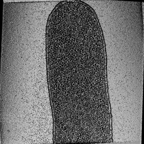
| タイトル | E coli cell with plasmid containing the ParMRC locus | |||||||||
 マップデータ マップデータ | E coli cell with plasmid containing the ParMRC locus | |||||||||
 試料 試料 |
| |||||||||
 キーワード キーワード | bacterial cytoskeleton / plasmid segregation / actin-like protein | |||||||||
| 生物種 |  | |||||||||
| 手法 | 電子線トモグラフィー法 / クライオ電子顕微鏡法 | |||||||||
 データ登録者 データ登録者 | Bharat TAM / Murshudov GN / Sachse C / Lowe J | |||||||||
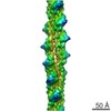
 引用 引用 | ジャーナル: Nature / 年: 2015 タイトル: Structures of actin-like ParM filaments show architecture of plasmid-segregating spindles. 著者: Tanmay A M Bharat / Garib N Murshudov / Carsten Sachse / Jan Löwe /   要旨: Active segregation of Escherichia coli low-copy-number plasmid R1 involves formation of a bipolar spindle made of left-handed double-helical actin-like ParM filaments. ParR links the filaments with ...Active segregation of Escherichia coli low-copy-number plasmid R1 involves formation of a bipolar spindle made of left-handed double-helical actin-like ParM filaments. ParR links the filaments with centromeric parC plasmid DNA, while facilitating the addition of subunits to ParM filaments. Growing ParMRC spindles push sister plasmids to the cell poles. Here, using modern electron cryomicroscopy methods, we investigate the structures and arrangements of ParM filaments in vitro and in cells, revealing at near-atomic resolution how subunits and filaments come together to produce the simplest known mitotic machinery. To understand the mechanism of dynamic instability, we determine structures of ParM filaments in different nucleotide states. The structure of filaments bound to the ATP analogue AMPPNP is determined at 4.3 Å resolution and refined. The ParM filament structure shows strong longitudinal interfaces and weaker lateral interactions. Also using electron cryomicroscopy, we reconstruct ParM doublets forming antiparallel spindles. Finally, with whole-cell electron cryotomography, we show that doublets are abundant in bacterial cells containing low-copy-number plasmids with the ParMRC locus, leading to an asynchronous model of R1 plasmid segregation. | |||||||||
| 履歴 |
|
- 構造の表示
構造の表示
| ムービー |
 ムービービューア ムービービューア |
|---|---|
| 構造ビューア | EMマップ: SurfViewMolmilJmol/JSmol |
| 添付画像 |
- ダウンロードとリンク
-EMDBアーカイブ
| マップデータ | emd_2849.map.gz | 625.8 MB | EMDBマップデータ形式 | |
|---|---|---|---|---|
| ヘッダ (付随情報) | emd-2849-v30.xmlemd-2849.xml | 8.1 KB 8.1 KB | 表示 表示 | EMDBヘッダ |
| 画像 |  emd-2849.png emd-2849.png | 431.5 KB | ||
| アーカイブディレクトリ |  http://ftp.pdbj.org/pub/emdb/structures/EMD-2849ftp://ftp.pdbj.org/pub/emdb/structures/EMD-2849 http://ftp.pdbj.org/pub/emdb/structures/EMD-2849ftp://ftp.pdbj.org/pub/emdb/structures/EMD-2849 | HTTPS FTP |
-関連構造データ


-リンク
| EMDBのページ | EMDB (EBI/PDBe) / EMDataResource |
|---|
-マップ
| ファイル | ダウンロード / ファイル: emd_2849.map.gz / 形式: CCP4 / 大きさ: 665.7 MB / タイプ: IMAGE STORED AS FLOATING POINT NUMBER (4 BYTES) | ||||||||||||||||||||||||||||||||||||||||||||||||||||||||||||
|---|---|---|---|---|---|---|---|---|---|---|---|---|---|---|---|---|---|---|---|---|---|---|---|---|---|---|---|---|---|---|---|---|---|---|---|---|---|---|---|---|---|---|---|---|---|---|---|---|---|---|---|---|---|---|---|---|---|---|---|---|---|
| 注釈 | E coli cell with plasmid containing the ParMRC locus | ||||||||||||||||||||||||||||||||||||||||||||||||||||||||||||
| 投影像・断面図 | 画像のコントロール
画像は Spider により作成 これらの図は立方格子座標系で作成されたものです | ||||||||||||||||||||||||||||||||||||||||||||||||||||||||||||
| ボクセルのサイズ | X=Y=Z: 17.8 Å | ||||||||||||||||||||||||||||||||||||||||||||||||||||||||||||
| 密度 |
| ||||||||||||||||||||||||||||||||||||||||||||||||||||||||||||
| 対称性 | 空間群: 1 | ||||||||||||||||||||||||||||||||||||||||||||||||||||||||||||
| 詳細 | EMDB XML:
CCP4マップ ヘッダ情報:
| ||||||||||||||||||||||||||||||||||||||||||||||||||||||||||||
 Z (Sec.)
 Y (Row.)
 X (Col.)


-添付データ
- 試料の構成要素
-全体 : E coli cell with plasmid containing medium copy number plasmid wi...
| 全体 | 名称: E coli cell with plasmid containing medium copy number plasmid with ParMRC locus |
|---|---|
| 要素 |
|
-超分子 #1000: E coli cell with plasmid containing medium copy number plasmid wi...
| 超分子 | 名称: E coli cell with plasmid containing medium copy number plasmid with ParMRC locus タイプ: sample / ID: 1000 / Number unique components: 1 |
|---|
-超分子 #1: Escherichia coli cytoskeleton
| 超分子 | 名称: Escherichia coli cytoskeleton / タイプ: organelle_or_cellular_component / ID: 1 / 詳細: 200 mesh copper / rhodium grid with carbon support / 組換発現: No / データベース: NCBI |
|---|---|
| 由来(天然) | 生物種: |
-実験情報
-構造解析
| 手法 | クライオ電子顕微鏡法 |
|---|---|
 解析 解析 | 電子線トモグラフィー法 |
| 試料の集合状態 | cell |
-試料調製
| グリッド | 詳細: 200 mesh copper / rhodium grid with carbon support |
|---|---|
| 凍結 | 凍結剤: ETHANE / チャンバー内湿度: 100 % / 装置: FEI VITROBOT MARK IV |
- 電子顕微鏡法
| 顕微鏡 | FEI TITAN KRIOS |
|---|---|
| 特殊光学系 | エネルギーフィルター - 名称: Gatan Quantum Energy Filter エネルギーフィルター - エネルギー下限: 0.0 eV エネルギーフィルター - エネルギー上限: 20.0 eV |
| 日付 | 2014年6月1日 |
| 撮影 | カテゴリ: CCD / フィルム・検出器のモデル: GATAN K2 (4k x 4k) / 実像数: 121 / 平均電子線量: 120 e/Å2 |
| 電子線 | 加速電圧: 300 kV / 電子線源:  FIELD EMISSION GUN FIELD EMISSION GUN |
| 電子光学系 | 照射モード: FLOOD BEAM / 撮影モード: BRIGHT FIELD / Cs: 2.7 mm / 最大 デフォーカス(公称値): -8.0 µm / 最小 デフォーカス(公称値): -8.0 µm |
| 試料ステージ | 試料ホルダーモデル: FEI TITAN KRIOS AUTOGRID HOLDER Tilt series - Axis1 - Angle increment: 1 ° |
| 実験機器 |  モデル: Titan Krios / 画像提供: FEI Company |
-画像解析
| 詳細 | Tilt series was aligned using IMOD and reconstructed using Tomo3D |
|---|---|
| 最終 再構成 | アルゴリズム: OTHER / ソフトウェア - 名称: IMOD, Tomo3D / 使用した粒子像数: 121 |
