 Movie
Movie Controller

[English] 日本語
 Yorodumi
- EMDB-2829: The interaction of an essential DNA helicase with RNA polymerase -
+ Open data

- Basic information
| Entry | Database: EMDB / ID: EMD-2829 | |||||||||
|---|---|---|---|---|---|---|---|---|---|---|
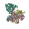
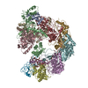
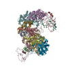
| Title | The interaction of an essential DNA helicase with RNA polymerase | |||||||||
 Map data Map data | Reconstruction of RNAP-PcrA complex | |||||||||
 Sample Sample |
| |||||||||
 Keywords Keywords | Transcription / RNA polymerase / helicase | |||||||||
| Biological species |  | |||||||||
| Method | single particle reconstruction / negative staining / Resolution: 25.0 Å | |||||||||
 Authors Authors | Harriott K / Yang X / Mervelet P / Buncherd H / Noirot P / de Jong L / Noirot Gros MF / Lewis PJ | |||||||||
 Citation Citation | Journal: To Be Published Title: The interaction of an essential DNA helicase with RNA polymerase Authors: Harriott K / Yang X / Mervelet P / Buncherd H / Noirot P / de Jong L / Noirot Gros MF / Lewis PJ | |||||||||
| History |
|
- Structure visualization
| Movie |
 Movie viewer Movie viewer |
|---|---|
| Structure viewer | EM map: SurfViewMolmilJmol/JSmol |
| Supplemental images |

- Downloads & links
-EMDB archive
| Map data | emd_2829.map.gz | 1.3 MB | EMDB map data format | |
|---|---|---|---|---|
| Header (meta data) | emd-2829-v30.xmlemd-2829.xml | 9 KB 9 KB | Display Display | EMDB header |
| Images | 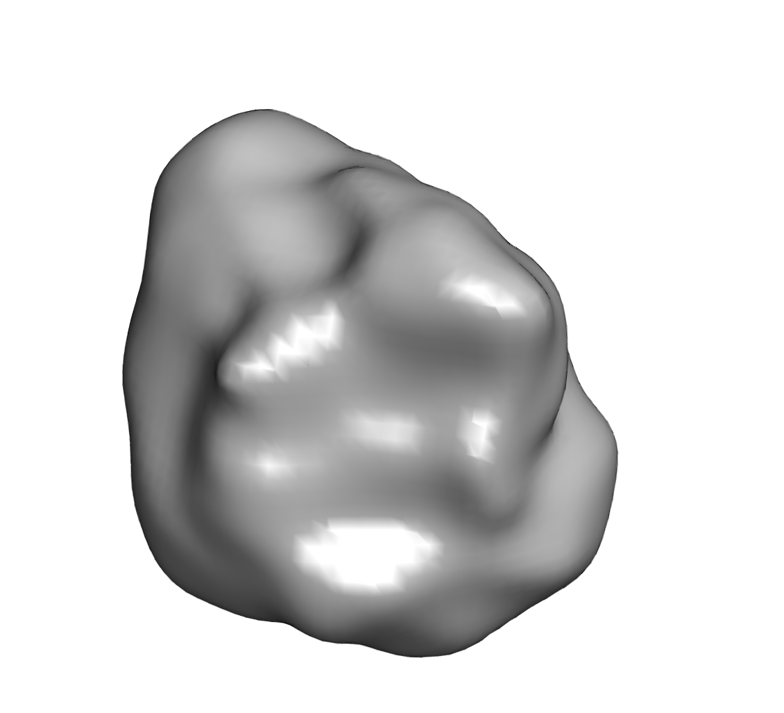 EMD-2829.png EMD-2829.png | 122.4 KB | ||
| Archive directory |  http://ftp.pdbj.org/pub/emdb/structures/EMD-2829ftp://ftp.pdbj.org/pub/emdb/structures/EMD-2829 http://ftp.pdbj.org/pub/emdb/structures/EMD-2829ftp://ftp.pdbj.org/pub/emdb/structures/EMD-2829 | HTTPS FTP |
-Validation report
| Summary document | emd_2829_validation.pdf.gz | 202.2 KB | Display | EMDB validaton report |
|---|---|---|---|---|
| Full document | emd_2829_full_validation.pdf.gz | 201.4 KB | Display | |
| Data in XML | emd_2829_validation.xml.gz | 5.3 KB | Display | |
| Arichive directory | https://ftp.pdbj.org/pub/emdb/validation_reports/EMD-2829ftp://ftp.pdbj.org/pub/emdb/validation_reports/EMD-2829 | HTTPS FTP |
-Related structure data
| Similar structure data |
|---|


-Links
| EMDB pages | EMDB (EBI/PDBe) / EMDataResource |
|---|
-Map
| File | Download / File: emd_2829.map.gz / Format: CCP4 / Size: 1.4 MB / Type: IMAGE STORED AS FLOATING POINT NUMBER (4 BYTES) | ||||||||||||||||||||||||||||||||||||||||||||||||||||||||||||||||||||
|---|---|---|---|---|---|---|---|---|---|---|---|---|---|---|---|---|---|---|---|---|---|---|---|---|---|---|---|---|---|---|---|---|---|---|---|---|---|---|---|---|---|---|---|---|---|---|---|---|---|---|---|---|---|---|---|---|---|---|---|---|---|---|---|---|---|---|---|---|---|
| Annotation | Reconstruction of RNAP-PcrA complex | ||||||||||||||||||||||||||||||||||||||||||||||||||||||||||||||||||||
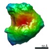
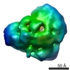
| Projections & slices | Image control
Images are generated by Spider. | ||||||||||||||||||||||||||||||||||||||||||||||||||||||||||||||||||||
| Voxel size | X=Y=Z: 3.9 Å | ||||||||||||||||||||||||||||||||||||||||||||||||||||||||||||||||||||
| Density |
| ||||||||||||||||||||||||||||||||||||||||||||||||||||||||||||||||||||
| Symmetry | Space group: 1 | ||||||||||||||||||||||||||||||||||||||||||||||||||||||||||||||||||||
| Details | EMDB XML:
CCP4 map header:
| ||||||||||||||||||||||||||||||||||||||||||||||||||||||||||||||||||||
 Z (Sec.)
 Y (Row.)
 X (Col.)


-Supplemental data
- Sample components
-Entire : RNAP-PcrA complex
| Entire | Name: RNAP-PcrA complex |
|---|---|
| Components |
|
-Supramolecule #1000: RNAP-PcrA complex
| Supramolecule | Name: RNAP-PcrA complex / type: sample / ID: 1000 / Details: complex was made with in vitro purified proteins / Oligomeric state: 1 / Number unique components: 2 |
|---|---|
| Molecular weight | Experimental: 370 KDa / Theoretical: 370 KDa |
-Macromolecule #1: RNAP
| Macromolecule | Name: RNAP / type: protein_or_peptide / ID: 1 / Number of copies: 1 / Oligomeric state: 1 / Recombinant expression: Yes |
|---|---|
| Source (natural) | Organism: |
| Molecular weight | Experimental: 370 KDa / Theoretical: 370 KDa |
| Recombinant expression | Organism: |
-Macromolecule #2: PcrA
| Macromolecule | Name: PcrA / type: protein_or_peptide / ID: 2 / Number of copies: 1 / Oligomeric state: 1 / Recombinant expression: Yes |
|---|---|
| Source (natural) | Organism: |
| Molecular weight | Experimental: 370 KDa / Theoretical: 370 KDa |
| Recombinant expression | Organism: |
-Experimental details
-Structure determination
| Method | negative staining |
|---|---|
 Processing Processing | single particle reconstruction |
| Aggregation state | particle |
-Sample preparation
| Concentration | 1 mg/mL |
|---|---|
| Buffer | pH: 7.8 / Details: 20 mM Tris, 150 mM NaCl, 10 mM MgCl2, 0.1 mM DTT |
| Staining | Type: NEGATIVE Details: Grids were stained with 1% uranyl formate for 30 seconds |
| Vitrification | Cryogen name: NONE / Instrument: OTHER |
- Electron microscopy
| Microscope | FEI TECNAI F30 |
|---|---|
| Date | Dec 3, 2008 |
| Image recording | Category: CCD / Film or detector model: GATAN K2 (4k x 4k) / Digitization - Sampling interval: 3.9 µm / Number real images: 240 / Bits/pixel: 16 |
| Electron beam | Acceleration voltage: 300 kV / Electron source:  FIELD EMISSION GUN FIELD EMISSION GUN |
| Electron optics | Illumination mode: FLOOD BEAM / Imaging mode: BRIGHT FIELD |
| Sample stage | Specimen holder: Eucentric / Specimen holder model: GATAN LIQUID NITROGEN |
| Experimental equipment |  Model: Tecnai F30 / Image courtesy: FEI Company |
-Image processing
| Final reconstruction | Applied symmetry - Point group: C1 (asymmetric) / Resolution.type: BY AUTHOR / Resolution: 25.0 Å / Resolution method: OTHER / Software - Name: EMAN / Number images used: 14266 |
|---|---|
| Final two d classification | Number classes: 1178 |
