ジャーナル: PNAS Nexus / 年: 2022 タイトル: Cryo-electron tomography with mixed-scale dense neural networks reveals key steps in deployment of invasion machinery. 著者: Li-Av Segev-Zarko / Peter D Dahlberg / Stella Y Sun / Daniël M Pelt / Chi Yong Kim / Elizabeth S Egan / James A Sethian / Wah Chiu / John C Boothroyd / 要旨: Host cell invasion by intracellular, eukaryotic parasites within the phylum Apicomplexa is a remarkable and active process involving the coordinated action of apical organelles and other structures. ...Host cell invasion by intracellular, eukaryotic parasites within the phylum Apicomplexa is a remarkable and active process involving the coordinated action of apical organelles and other structures. To date, capturing how these structures interact during invasion has been difficult to observe in detail. Here, we used cryogenic electron tomography to image the apical complex of tachyzoites under conditions that mimic resting parasites and those primed to invade through stimulation with calcium ionophore. Through the application of mixed-scale dense networks for image processing, we developed a highly efficient pipeline for annotation of tomograms, enabling us to identify and extract densities of relevant subcellular organelles and accurately analyze features in 3-D. The results reveal a dramatic change in the shape of the anteriorly located apical vesicle upon its apparent fusion with a rhoptry that occurs only in the stimulated parasites. We also present information indicating that this vesicle originates from the vesicles that parallel the intraconoidal microtubules and that the latter two structures are linked by a novel tether. We show that a rosette structure previously proposed to be involved in rhoptry secretion is associated with apical vesicles beyond just the most anterior one. This result, suggesting multiple vesicles are primed to enable rhoptry secretion, may shed light on the mechanisms employs to enable repeated invasion attempts. Using the same approach, we examine merozoites and show that they too possess an apical vesicle just beneath a rosette, demonstrating evolutionary conservation of this overall subcellular organization.
A: 13286.4 Å / B: 12843.5205 Å / C: 2892.56 Å α=β=γ: 90.0 °
-
添付データ
-
試料の構成要素
-
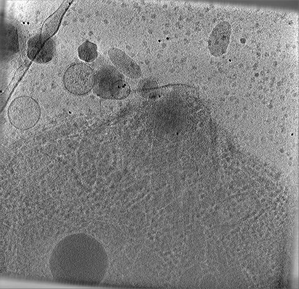
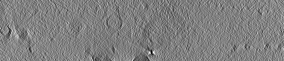
全体 : The apical complex of Toxoplasma gondii (non-stimulated)
全体
名称: The apical complex of Toxoplasma gondii (non-stimulated)
要素
細胞器官・細胞要素: The apical complex of Toxoplasma gondii (non-stimulated)
-
超分子 #1: The apical complex of Toxoplasma gondii (non-stimulated)
超分子
名称: The apical complex of Toxoplasma gondii (non-stimulated) タイプ: organelle_or_cellular_component / ID: 1 / 親要素: 0
由来(天然)
生物種: Toxoplasma gondii (トキソプラズマ) / 株: RH
-
実験情報
-
構造解析
手法
クライオ電子顕微鏡法
解析
電子線トモグラフィー法
試料の集合状態
cell
-
試料調製
緩衝液
pH: 7.2 / 構成要素 - 濃度: 1.0 x / 構成要素 - 名称: Endo buffer 詳細: 45 mM potassium sulfate, 106 mM sucrose, 10 mM magnesium sulfate, 20 mM Tris buffer pH 7.2, 5 mM glucose and 0.35% bovine serum albumin
凍結剤: ETHANE / チャンバー内湿度: 90 % / チャンバー内温度: 297 K / 装置: LEICA EM GP
詳細
Tachyzoites were released from heavily infected monolayers of HFFs by mechanical disruption of the monolayers using disposable scrapers and passage through a 25-gauge syringe into HPEB
切片作成
その他: NO SECTIONING
位置合わせマーカー
Manufacturer: EMS / 直径: 10 nm
-
電子顕微鏡法
顕微鏡
TFS KRIOS
特殊光学系
位相板: VOLTA PHASE PLATE / エネルギーフィルター - 名称: GIF Bioquantum
撮影
フィルム・検出器のモデル: GATAN K2 SUMMIT (4k x 4k) 平均電子線量: 1.0 e/Å2
 ムービー
ムービー コントローラー


 データを開く
 基本情報
基本情報
 マップデータ
 試料
試料 キーワード
キーワード
 データ登録者
 米国, 2件
  引用
引用
 構造の表示
構造の表示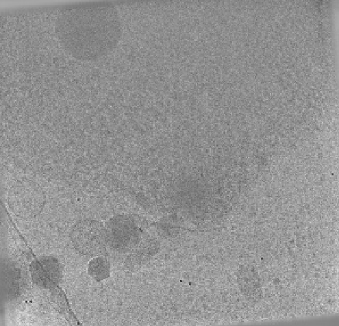
 ダウンロードとリンク
 EMDBマップデータ形式
 emd_28140.png
 http://ftp.pdbj.org/pub/emdb/structures/EMD-28140
 Z (Sec.)
 Y (Row.)
 X (Col.)


 試料の構成要素
 解析
解析 電子顕微鏡法
 FIELD EMISSION GUN
