National Institutes of Health/National Institute of General Medical Sciences (NIH/NIGMS)
R01GM079429
米国
National Institutes of Health/National Institute Of Allergy and Infectious Diseases (NIH/NIAID)
DP2HL137186
米国
引用
ジャーナル: mBio / 年: 2024 タイトル: Cryogenic electron tomography reveals novel structures in the apical complex of . 著者: Stella Y Sun / Li-Av Segev-Zarko / Grigore D Pintilie / Chi Yong Kim / Sophia R Staggers / Michael F Schmid / Elizabeth S Egan / Wah Chiu / John C Boothroyd / 要旨: Intracellular infectious agents, like the malaria parasite, , face the daunting challenge of how to invade a host cell. This problem may be even harder when the host cell in question is the ...Intracellular infectious agents, like the malaria parasite, , face the daunting challenge of how to invade a host cell. This problem may be even harder when the host cell in question is the enucleated red blood cell, which lacks the host machinery co-opted by many pathogens for internalization. Evolution has provided and related single-celled parasites within the phylum Apicomplexa with a collection of organelles at their apical end that mediate invasion. This apical complex includes at least two sets of secretory organelles, micronemes and rhoptries, and several structural features like apical rings and a putative pore through which proteins may be introduced into the host cell during invasion. We perform cryogenic electron tomography (cryo-ET) equipped with Volta Phase Plate on isolated and vitrified merozoites to visualize the apical machinery. Through tomographic reconstruction of cellular compartments, we see new details of known structures like the rhoptry tip interacting directly with a rosette resembling the recently described rhoptry secretory apparatus (RSA) or with an apical vesicle docked beneath the RSA. Subtomogram averaging reveals that the apical rings have a fixed number of repeating units, each of which is similar in overall size and shape to the units in the apical rings of tachyzoites of . Comparison of these polar rings in and parasites also reveals them to have a structurally conserved assembly pattern. These results provide new insight into the essential and structurally conserved features of this remarkable machinery used by apicomplexan parasites to invade their respective host cells. IMPORTANCE: Malaria is an infectious disease caused by parasites of the genus and is a leading cause of morbidity and mortality globally. Upon infection, parasites invade and replicate in red blood ...IMPORTANCE: Malaria is an infectious disease caused by parasites of the genus and is a leading cause of morbidity and mortality globally. Upon infection, parasites invade and replicate in red blood cells, where they are largely protected from the immune system. To enter host cells, the parasites employ a specialized apparatus at their anterior end. In this study, advanced imaging techniques like cryogenic electron tomography (cryo-ET) and Volta Phase Plate enable unprecedented visualization of whole merozoites, revealing previously unknown structural details of their invasion machinery. Key findings include new insights into the structural conservation of apical rings shared between and its apicomplexan cousin, . These discoveries shed light on the essential and conserved elements of the invasion machinery used by these pathogens. Moreover, the research provides a foundation for understanding the molecular mechanisms underlying parasite-host interactions, potentially informing strategies for combating diseases caused by apicomplexan parasites.
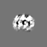
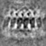
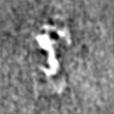
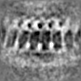
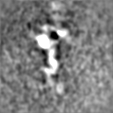
This map is obtained by positioning the reconstructed full map at multiple positions around the rings in tomograms. This map is cropped to show a small segment of the entire rings.
 ムービー
ムービー コントローラー


 データを開く
 基本情報
基本情報
 マップデータ
 試料
試料 キーワード
キーワード
 データ登録者
 米国, 2件
  引用
引用 構造の表示
構造の表示
 ダウンロードとリンク
 EMDBマップデータ形式
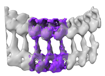 emd_28126.png
 http://ftp.pdbj.org/pub/emdb/structures/EMD-28126

 Z (Sec.)
 Y (Row.)
 X (Col.)


 試料の構成要素
 解析
解析 電子顕微鏡法
 FIELD EMISSION GUN
