Movie
Movie Controller


+ Open data

- Basic information
| Entry |  | ||||||||||||
|---|---|---|---|---|---|---|---|---|---|---|---|---|---|
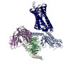
| Title | Morphine-bound mu-opioid receptor-Gi complex | ||||||||||||
 Map data Map data | |||||||||||||
 Sample Sample |
| ||||||||||||
 Keywords Keywords | mu-opioid receptor / G protein / morphine / SIGNALING PROTEIN | ||||||||||||
| Function / homology |  Function and homology information Function and homology informationOpioid Signalling / beta-endorphin receptor activity / morphine receptor activity / negative regulation of Wnt protein secretion / regulation of cellular response to stress / G protein-coupled opioid receptor signaling pathway / negative regulation of nitric oxide biosynthetic process / adenylate cyclase-inhibiting G protein-coupled acetylcholine receptor signaling pathway / behavioral response to ethanol / G-protein activation ...Opioid Signalling / beta-endorphin receptor activity / morphine receptor activity / negative regulation of Wnt protein secretion / regulation of cellular response to stress / G protein-coupled opioid receptor signaling pathway / negative regulation of nitric oxide biosynthetic process / adenylate cyclase-inhibiting G protein-coupled acetylcholine receptor signaling pathway / behavioral response to ethanol / G-protein activation / Activation of the phototransduction cascade / Glucagon-type ligand receptors / Thromboxane signalling through TP receptor / Sensory perception of sweet, bitter, and umami (glutamate) taste / G beta:gamma signalling through PI3Kgamma / G beta:gamma signalling through CDC42 / Cooperation of PDCL (PhLP1) and TRiC/CCT in G-protein beta folding / Activation of G protein gated Potassium channels / Inhibition of voltage gated Ca2+ channels via Gbeta/gamma subunits / Ca2+ pathway / G alpha (z) signalling events / High laminar flow shear stress activates signaling by PIEZO1 and PECAM1:CDH5:KDR in endothelial cells / Glucagon-like Peptide-1 (GLP1) regulates insulin secretion / Vasopressin regulates renal water homeostasis via Aquaporins / Adrenaline,noradrenaline inhibits insulin secretion / ADP signalling through P2Y purinoceptor 12 / regulation of NMDA receptor activity / G alpha (q) signalling events / G alpha (i) signalling events / Thrombin signalling through proteinase activated receptors (PARs) / Activation of G protein gated Potassium channels / G-protein activation / G beta:gamma signalling through PI3Kgamma / Prostacyclin signalling through prostacyclin receptor / G beta:gamma signalling through PLC beta / ADP signalling through P2Y purinoceptor 1 / Thromboxane signalling through TP receptor / Presynaptic function of Kainate receptors / G beta:gamma signalling through CDC42 / Inhibition of voltage gated Ca2+ channels via Gbeta/gamma subunits / neuropeptide binding / G alpha (12/13) signalling events / Glucagon-type ligand receptors / G beta:gamma signalling through BTK / ADP signalling through P2Y purinoceptor 12 / Adrenaline,noradrenaline inhibits insulin secretion / Cooperation of PDCL (PhLP1) and TRiC/CCT in G-protein beta folding / Ca2+ pathway / G alpha (z) signalling events / Thrombin signalling through proteinase activated receptors (PARs) / positive regulation of neurogenesis / Extra-nuclear estrogen signaling / G alpha (s) signalling events / G alpha (q) signalling events / sensory perception / photoreceptor outer segment membrane / spectrin binding / Glucagon-like Peptide-1 (GLP1) regulates insulin secretion / G alpha (i) signalling events / High laminar flow shear stress activates signaling by PIEZO1 and PECAM1:CDH5:KDR in endothelial cells / Vasopressin regulates renal water homeostasis via Aquaporins / negative regulation of cytosolic calcium ion concentration / alkylglycerophosphoethanolamine phosphodiesterase activity / photoreceptor outer segment / G-protein alpha-subunit binding / G protein-coupled receptor signaling pathway, coupled to cyclic nucleotide second messenger / voltage-gated calcium channel activity / MECP2 regulates neuronal receptors and channels / regulation of eating behavior / cardiac muscle cell apoptotic process / adenylate cyclase inhibitor activity / photoreceptor inner segment / positive regulation of protein localization to cell cortex / T cell migration / positive regulation of relaxation of smooth muscle / Adenylate cyclase inhibitory pathway / D2 dopamine receptor binding / sensory perception of pain / adenylate cyclase-inhibiting serotonin receptor signaling pathway / G protein-coupled serotonin receptor binding / cellular response to forskolin / mast cell degranulation / Peptide ligand-binding receptors / regulation of mitotic spindle organization / chemokine-mediated signaling pathway / Regulation of insulin secretion / neuropeptide signaling pathway / response to prostaglandin E / positive regulation of cholesterol biosynthetic process / G protein-coupled receptor binding / response to peptide hormone / G protein-coupled receptor activity / G-protein beta/gamma-subunit complex binding / adenylate cyclase-modulating G protein-coupled receptor signaling pathway / adenylate cyclase-inhibiting G protein-coupled receptor signaling pathway / G-protein activation / GDP binding / ADP signalling through P2Y purinoceptor 12 / Adrenaline,noradrenaline inhibits insulin secretion / G alpha (z) signalling events Similarity search - Function | ||||||||||||
| Biological species |  Homo sapiens (human) / Homo sapiens (human) /  | ||||||||||||
| Method | single particle reconstruction / cryo EM / Resolution: 3.2 Å | ||||||||||||
 Authors Authors | Zhuang Y / Wang Y / Guo S / Zhou XE / Rao Q / He X / He B / Liu J / Zhou Q / Wang X ...Zhuang Y / Wang Y / Guo S / Zhou XE / Rao Q / He X / He B / Liu J / Zhou Q / Wang X / Liu W / Jiang X / Yang D / Chen X / Jiang Y / Jiang H / Shen J / Melcher K / Wang M / Xie X / Xu HE | ||||||||||||
| Funding support |  China, 3 items China, 3 items
| ||||||||||||
 Citation Citation | Journal: Cell / Year: 2022 Title: Molecular recognition of morphine and fentanyl by the human μ-opioid receptor. Authors: Youwen Zhuang / Yue Wang / Bingqing He / Xinheng He / X Edward Zhou / Shimeng Guo / Qidi Rao / Jiaqi Yang / Jinyu Liu / Qingtong Zhou / Xiaoxi Wang / Mingliang Liu / Weiyi Liu / Xiangrui ...Authors: Youwen Zhuang / Yue Wang / Bingqing He / Xinheng He / X Edward Zhou / Shimeng Guo / Qidi Rao / Jiaqi Yang / Jinyu Liu / Qingtong Zhou / Xiaoxi Wang / Mingliang Liu / Weiyi Liu / Xiangrui Jiang / Dehua Yang / Hualiang Jiang / Jingshan Shen / Karsten Melcher / Hong Chen / Yi Jiang / Xi Cheng / Ming-Wei Wang / Xin Xie / H Eric Xu /  Abstract: Morphine and fentanyl are among the most used opioid drugs that confer analgesia and unwanted side effects through both G protein and arrestin signaling pathways of μ-opioid receptor (μOR). Here, ...Morphine and fentanyl are among the most used opioid drugs that confer analgesia and unwanted side effects through both G protein and arrestin signaling pathways of μ-opioid receptor (μOR). Here, we report structures of the human μOR-G protein complexes bound to morphine and fentanyl, which uncover key differences in how they bind the receptor. We also report structures of μOR bound to TRV130, PZM21, and SR17018, which reveal preferential interactions of these agonists with TM3 side of the ligand-binding pocket rather than TM6/7 side. In contrast, morphine and fentanyl form dual interactions with both TM3 and TM6/7 regions. Mutations at the TM6/7 interface abolish arrestin recruitment of μOR promoted by morphine and fentanyl. Ligands designed to reduce TM6/7 interactions display preferential G protein signaling. Our results provide crucial insights into fentanyl recognition and signaling of μOR, which may facilitate rational design of next-generation analgesics. | ||||||||||||
| History |
|
- Structure visualization
| Supplemental images |
|---|

- Downloads & links
-EMDB archive
| Map data | emd_28069.map.gz | 38 MB | EMDB map data format | |
|---|---|---|---|---|
| Header (meta data) | emd-28069-v30.xmlemd-28069.xml | 25.3 KB 25.3 KB | Display Display | EMDB header |
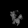
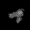
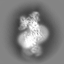
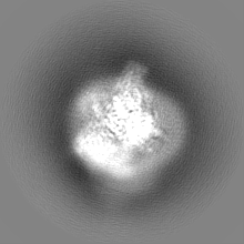
| Images | 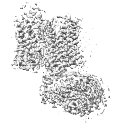 emd_28069.png emd_28069.png | 117.2 KB | ||
| Filedesc metadata | emd-28069.cif.gz | 7.5 KB | ||
| Others | emd_28069_half_map_1.map.gzemd_28069_half_map_2.map.gz | 31.3 MB 31.3 MB | ||
| Archive directory |  http://ftp.pdbj.org/pub/emdb/structures/EMD-28069ftp://ftp.pdbj.org/pub/emdb/structures/EMD-28069 http://ftp.pdbj.org/pub/emdb/structures/EMD-28069ftp://ftp.pdbj.org/pub/emdb/structures/EMD-28069 | HTTPS FTP |
-Related structure data
| Related structure data |  8ef6MC  8ef5C  8efbC  8eflC  8efoC  8efqC C: citing same article ( M: atomic model generated by this map |
|---|---|
| Similar structure data |
-Links
| EMDB pages | EMDB (EBI/PDBe) / EMDataResource |
|---|---|
| Related items in Molecule of the Month |


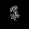
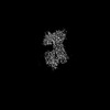
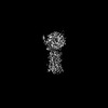
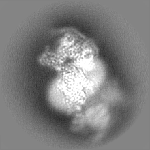
-Map
| File | Download / File: emd_28069.map.gz / Format: CCP4 / Size: 40.6 MB / Type: IMAGE STORED AS FLOATING POINT NUMBER (4 BYTES) | ||||||||||||||||||||||||||||||||||||
|---|---|---|---|---|---|---|---|---|---|---|---|---|---|---|---|---|---|---|---|---|---|---|---|---|---|---|---|---|---|---|---|---|---|---|---|---|---|
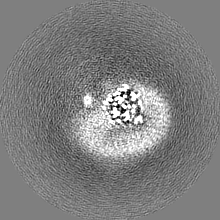
| Projections & slices | Image control
Images are generated by Spider. | ||||||||||||||||||||||||||||||||||||
| Voxel size | X=Y=Z: 1.071 Å | ||||||||||||||||||||||||||||||||||||
| Density |
| ||||||||||||||||||||||||||||||||||||
| Symmetry | Space group: 1 | ||||||||||||||||||||||||||||||||||||
| Details | EMDB XML:
|
 Z (Sec.)
 Y (Row.)
 X (Col.)


-Supplemental data
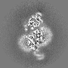
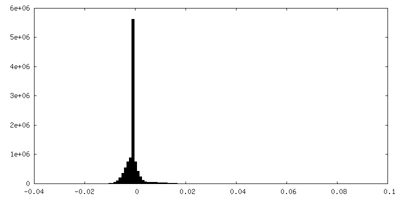
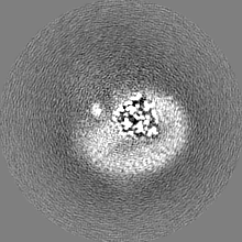
-Half map: #1
| File | emd_28069_half_map_1.map | ||||||||||||
|---|---|---|---|---|---|---|---|---|---|---|---|---|---|
| Projections & Slices |
| ||||||||||||
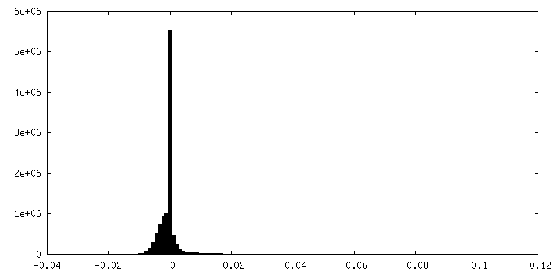
| Density Histograms |


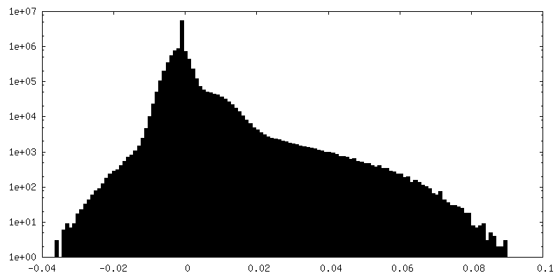
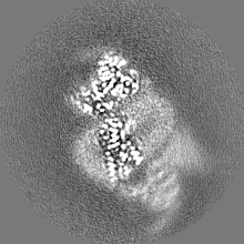
-Half map: #2
| File | emd_28069_half_map_2.map | ||||||||||||
|---|---|---|---|---|---|---|---|---|---|---|---|---|---|
| Projections & Slices |
| ||||||||||||
| Density Histograms |


- Sample components
-Entire : fentanyl bound mu-opioid receptor-G protein complex
| Entire | Name: fentanyl bound mu-opioid receptor-G protein complex |
|---|---|
| Components |
|
-Supramolecule #1: fentanyl bound mu-opioid receptor-G protein complex
| Supramolecule | Name: fentanyl bound mu-opioid receptor-G protein complex / type: complex / ID: 1 / Parent: 0 / Macromolecule list: #1-#5 |
|---|---|
| Source (natural) | Organism: Homo sapiens (human) |
-Macromolecule #1: Mu-type opioid receptor
| Macromolecule | Name: Mu-type opioid receptor / type: protein_or_peptide / ID: 1 / Number of copies: 2 / Enantiomer: LEVO |
|---|---|
| Source (natural) | Organism: Homo sapiens (human) |
| Molecular weight | Theoretical: 41.072191 KDa |
| Recombinant expression | Organism:   Spodoptera frugiperda (fall armyworm) Spodoptera frugiperda (fall armyworm) |
| Sequence | String: DSSAAPTNAS NCTDALAYSS CSPAPSPGSW VNLSHLDGNL SDPCGPNRTD LGGRDSLCPP TGSPSMITAI TIMALYSIVC VVGLFGNFL VMYVIVRYTK MKTATNIYIF NLALADALAT STLPFQSVNY LMGTWPFGTI LCKIVISIDY YNMFTSIFTL C TMSVDRYI ...String: DSSAAPTNAS NCTDALAYSS CSPAPSPGSW VNLSHLDGNL SDPCGPNRTD LGGRDSLCPP TGSPSMITAI TIMALYSIVC VVGLFGNFL VMYVIVRYTK MKTATNIYIF NLALADALAT STLPFQSVNY LMGTWPFGTI LCKIVISIDY YNMFTSIFTL C TMSVDRYI AVCHPVKALD FRTPRNAKII NVCNWILSSA IGLPVMFMAT TKYRQGSIDC TLTFSHPTWY WENLLKICVF IF AFIMPVL IITVCYGLMI LRLKSVRMLS GSKEKDRNLR RITRMVLVVV AVFIVCWTPI HIYVIIKALV TIPETTFQTV SWH FCIALG YTNSCLNPVL YAFLDENFKR CFREFCIPTS SNIEQQNSTR I UniProtKB: Mu-type opioid receptor |
-Macromolecule #2: Guanine nucleotide-binding protein G(i) subunit alpha-1
| Macromolecule | Name: Guanine nucleotide-binding protein G(i) subunit alpha-1 type: protein_or_peptide / ID: 2 / Number of copies: 2 / Enantiomer: LEVO |
|---|---|
| Source (natural) | Organism: Homo sapiens (human) |
| Molecular weight | Theoretical: 40.445059 KDa |
| Recombinant expression | Organism: Spodoptera frugiperda (fall armyworm) |
| Sequence | String: MGCTLSAEDK AAVERSKMID RNLREDGEKA AREVKLLLLG AGESGKSTIV KQMKIIHEAG YSEEECKQYK AVVYSNTIQS IIAIIRAMG RLKIDFGDSA RADDARQLFV LAGAAEEGFM TAELAGVIKR LWKDSGVQAC FNRSREYQLN DSAAYYLNDL D RIAQPNYI ...String: MGCTLSAEDK AAVERSKMID RNLREDGEKA AREVKLLLLG AGESGKSTIV KQMKIIHEAG YSEEECKQYK AVVYSNTIQS IIAIIRAMG RLKIDFGDSA RADDARQLFV LAGAAEEGFM TAELAGVIKR LWKDSGVQAC FNRSREYQLN DSAAYYLNDL D RIAQPNYI PTQQDVLRTR VKTTGIVETH FTFKDLHFKM FDVGAQRSER KKWIHCFEGV TAIIFCVALS DYDLVLAEDE EM NRMHESM KLFDSICNNK WFTDTSIILF LNKKDLFEEK IKKSPLTICY PEYAGSNTYE EAAAYIQCQF EDLNKRKDTK EIY THFTCS TDTKNVQFVF DAVTDVIIKN NLKDCGLF UniProtKB: Guanine nucleotide-binding protein G(i) subunit alpha-1 |
-Macromolecule #3: Guanine nucleotide-binding protein G(I)/G(S)/G(T) subunit beta-1
| Macromolecule | Name: Guanine nucleotide-binding protein G(I)/G(S)/G(T) subunit beta-1 type: protein_or_peptide / ID: 3 / Number of copies: 1 / Enantiomer: LEVO |
|---|---|
| Source (natural) | Organism: |
| Molecular weight | Theoretical: 39.020664 KDa |
| Recombinant expression | Organism: Spodoptera frugiperda (fall armyworm) |
| Sequence | String: MHHHHHHHHG SLLQSELDQL RQEAEQLKNQ IRDARKACAD ATLSQITNNI DPVGRIQMRT RRTLRGHLAK IYAMHWGTDS RLLVSASQD GKLIIWDSYT TNKVHAIPLR SSWVMTCAYA PSGNYVACGG LDNICSIYNL KTREGNVRVS RELAGHTGYL S CCRFLDDN ...String: MHHHHHHHHG SLLQSELDQL RQEAEQLKNQ IRDARKACAD ATLSQITNNI DPVGRIQMRT RRTLRGHLAK IYAMHWGTDS RLLVSASQD GKLIIWDSYT TNKVHAIPLR SSWVMTCAYA PSGNYVACGG LDNICSIYNL KTREGNVRVS RELAGHTGYL S CCRFLDDN QIVTSSGDTT CALWDIETGQ QTTTFTGHTG DVMSLSLAPD TRLFVSGACD ASAKLWDVRE GMCRQTFTGH ES DINAICF FPNGNAFATG SDDATCRLFD LRADQELMTY SHDNIICGIT SVSFSKSGRL LLAGYDDFNC NVWDALKADR AGV LAGHDN RVSCLGVTDD GMAVATGSWD SFLKIWN UniProtKB: Guanine nucleotide-binding protein G(I)/G(S)/G(T) subunit beta-1 |
-Macromolecule #4: Guanine nucleotide-binding protein G(I)/G(S)/G(O) subunit gamma-2
| Macromolecule | Name: Guanine nucleotide-binding protein G(I)/G(S)/G(O) subunit gamma-2 type: protein_or_peptide / ID: 4 / Number of copies: 1 / Enantiomer: LEVO |
|---|---|
| Source (natural) | Organism: |
| Molecular weight | Theoretical: 7.56375 KDa |
| Recombinant expression | Organism: Spodoptera frugiperda (fall armyworm) |
| Sequence | String: MASNNTASIA QARKLVEQLK MEANIDRIKV SKAAADLMAY CEAHAKEDPL LTPVPASENP FREKKFFC UniProtKB: Guanine nucleotide-binding protein G(I)/G(S)/G(O) subunit gamma-2 |
-Macromolecule #5: scFV16
| Macromolecule | Name: scFV16 / type: protein_or_peptide / ID: 5 / Number of copies: 1 / Enantiomer: LEVO |
|---|---|
| Source (natural) | Organism: synthetic construct (others) |
| Molecular weight | Theoretical: 26.408492 KDa |
| Recombinant expression | Organism:  |
| Sequence | String: MVQLVESGGG LVQPGGSRKL SCSASGFAFS SFGMHWVRQA PEKGLEWVAY ISSGSGTIYY ADTVKGRFTI SRDDPKNTLF LQMTSLRSE DTAMYYCVRS IYYYGSSPFD FWGQGTTLTV SAGGGGSGGG GSGGGGSADI VMTQATSSVP VTPGESVSIS C RSSKSLLH ...String: MVQLVESGGG LVQPGGSRKL SCSASGFAFS SFGMHWVRQA PEKGLEWVAY ISSGSGTIYY ADTVKGRFTI SRDDPKNTLF LQMTSLRSE DTAMYYCVRS IYYYGSSPFD FWGQGTTLTV SAGGGGSGGG GSGGGGSADI VMTQATSSVP VTPGESVSIS C RSSKSLLH SNGNTYLYWF LQRPGQSPQL LIYRMSNLAS GVPDRFSGSG SGTAFTLTIS RLEAEDVGVY YCMQHLEYPL TF GAGTKLE L |
-Macromolecule #6: (7R,7AS,12BS)-3-METHYL-2,3,4,4A,7,7A-HEXAHYDRO-1H-4,12-METHANO[1]...
| Macromolecule | Name: (7R,7AS,12BS)-3-METHYL-2,3,4,4A,7,7A-HEXAHYDRO-1H-4,12-METHANO[1]BENZOFURO[3,2-E]ISOQUINOLINE-7,9-DIOL type: ligand / ID: 6 / Number of copies: 2 / Formula: MOI |
|---|---|
| Molecular weight | Theoretical: 285.338 Da |
| Chemical component information | 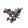 ChemComp-MOI: |
-Macromolecule #7: CHOLESTEROL
| Macromolecule | Name: CHOLESTEROL / type: ligand / ID: 7 / Number of copies: 10 / Formula: CLR |
|---|---|
| Molecular weight | Theoretical: 386.654 Da |
| Chemical component information |  ChemComp-CLR: |
-Experimental details
-Structure determination
| Method | cryo EM |
|---|---|
 Processing Processing | single particle reconstruction |
| Aggregation state | particle |
-Sample preparation
| Buffer | pH: 7.2 |
|---|---|
| Vitrification | Cryogen name: ETHANE |
- Electron microscopy
| Microscope | FEI TITAN KRIOS |
|---|---|
| Image recording | Film or detector model: GATAN K3 (6k x 4k) / Average electron dose: 70.0 e/Å2 |
| Electron beam | Acceleration voltage: 300 kV / Electron source:  FIELD EMISSION GUN FIELD EMISSION GUN |
| Electron optics | Illumination mode: FLOOD BEAM / Imaging mode: BRIGHT FIELD / Nominal defocus max: 5.0 µm / Nominal defocus min: 3.0 µm |
| Experimental equipment |  Model: Titan Krios / Image courtesy: FEI Company |