National Institutes of Health/National Eye Institute (NIH/NEI)
5F31EY030763
United States
National Institutes of Health/National Institute of General Medical Sciences (NIH/NIGMS)
5R01GM127652
United States
Citation
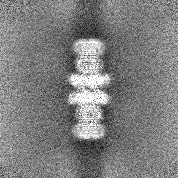
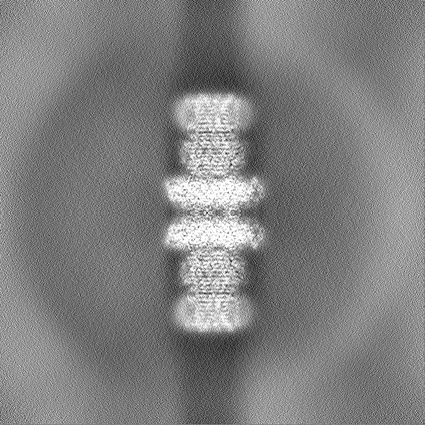
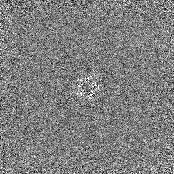
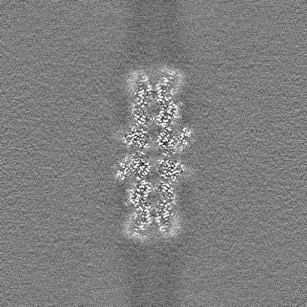
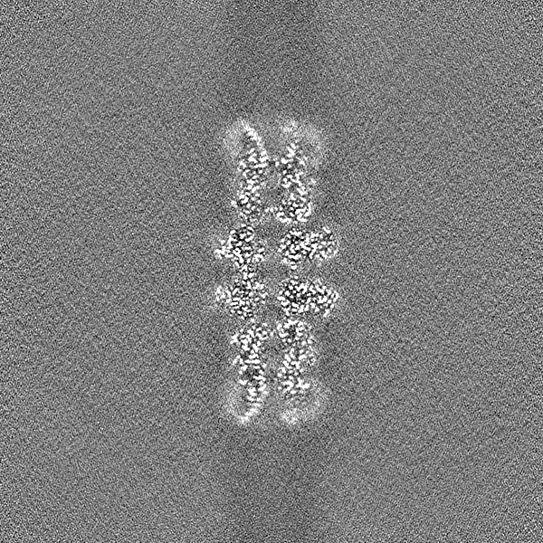
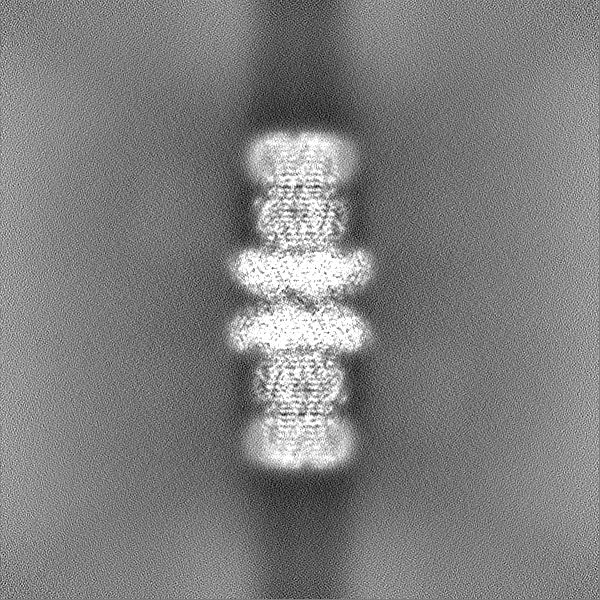
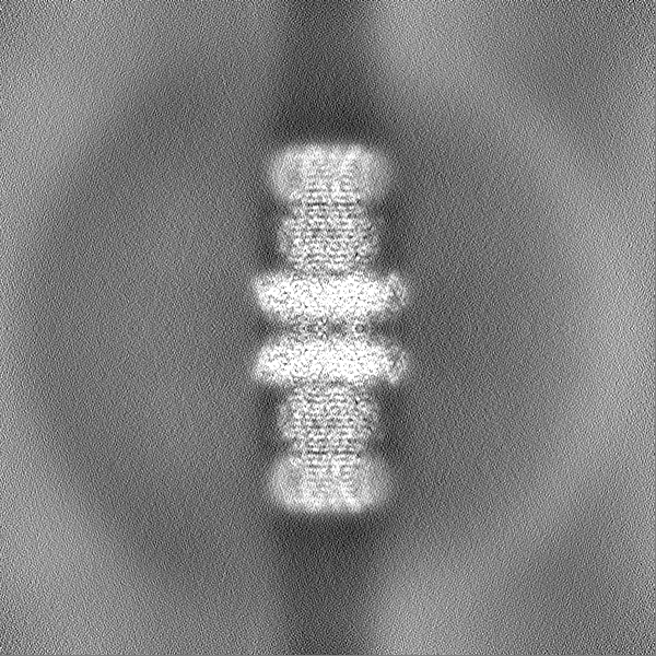
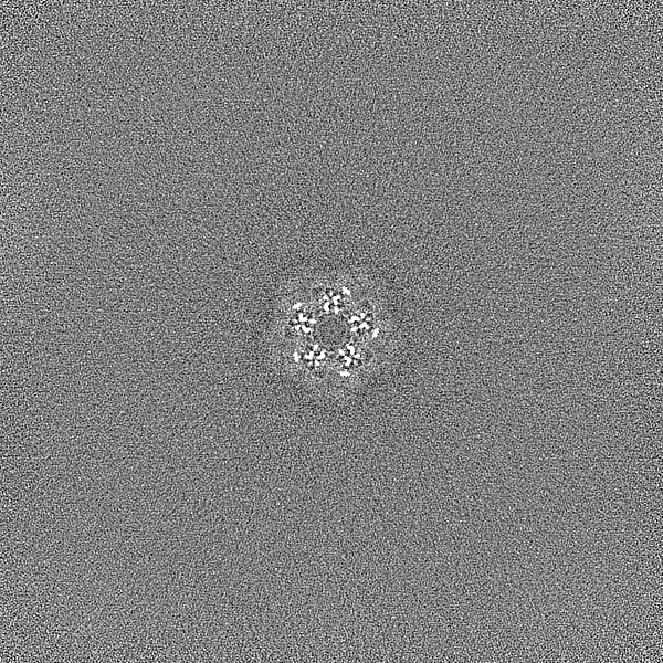
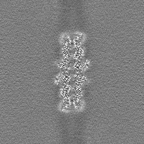
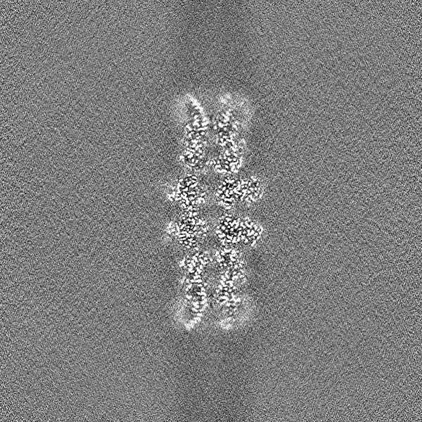
Journal: Nature / Year: 2022 Title: Bestrophin-2 and glutamine synthetase form a complex for glutamate release. Authors: Aaron P Owji / Kuai Yu / Alec Kittredge / Jiali Wang / Yu Zhang / Tingting Yang / Abstract: Bestrophin-2 (BEST2) is a member of the bestrophin family of calcium-activated anion channels that has a critical role in ocular physiology. Here we uncover a directional permeability of BEST2 to ...Bestrophin-2 (BEST2) is a member of the bestrophin family of calcium-activated anion channels that has a critical role in ocular physiology. Here we uncover a directional permeability of BEST2 to glutamate that heavily favours glutamate exit, identify glutamine synthetase (GS) as a binding partner of BEST2 in the ciliary body of the eye, and solve the structure of the BEST2-GS complex. BEST2 reduces cytosolic GS activity by tethering GS to the cell membrane. GS extends the ion conducting pathway of BEST2 through its central cavity and inhibits BEST2 channel function in the absence of intracellular glutamate, but sensitizes BEST2 to intracellular glutamate, which promotes the opening of BEST2 and thus relieves the inhibitory effect of GS. We demonstrate the physiological role of BEST2 in conducting chloride and glutamate and the influence of GS in non-pigmented ciliary epithelial cells. Together, our results reveal a novel mechanism of glutamate release through BEST2-GS.
In the structure databanks used in Yorodumi, some data are registered as the other names, "COVID-19 virus" and "2019-nCoV". Here are the details of the virus and the list of structure data.
Jan 31, 2019. EMDB accession codes are about to change! (news from PDBe EMDB page)
EMDB accession codes are about to change! (news from PDBe EMDB page)
The allocation of 4 digits for EMDB accession codes will soon come to an end. Whilst these codes will remain in use, new EMDB accession codes will include an additional digit and will expand incrementally as the available range of codes is exhausted. The current 4-digit format prefixed with “EMD-” (i.e. EMD-XXXX) will advance to a 5-digit format (i.e. EMD-XXXXX), and so on. It is currently estimated that the 4-digit codes will be depleted around Spring 2019, at which point the 5-digit format will come into force.
The EM Navigator/Yorodumi systems omit the EMD- prefix.
Related info.:Q: What is EMD? / ID/Accession-code notation in Yorodumi/EM Navigator
Yorodumi is a browser for structure data from EMDB, PDB, SASBDB, etc.
This page is also the successor to EM Navigator detail page, and also detail information page/front-end page for Omokage search.
The word "yorodu" (or yorozu) is an old Japanese word meaning "ten thousand". "mi" (miru) is to see.
Related info.:EMDB / PDB / SASBDB / Comparison of 3 databanks / Yorodumi Search / Aug 31, 2016. New EM Navigator & Yorodumi / Yorodumi Papers / Jmol/JSmol / Function and homology information / Changes in new EM Navigator and Yorodumi
 Movie
Movie Controller

 Yorodumi
 Open data
 Basic information
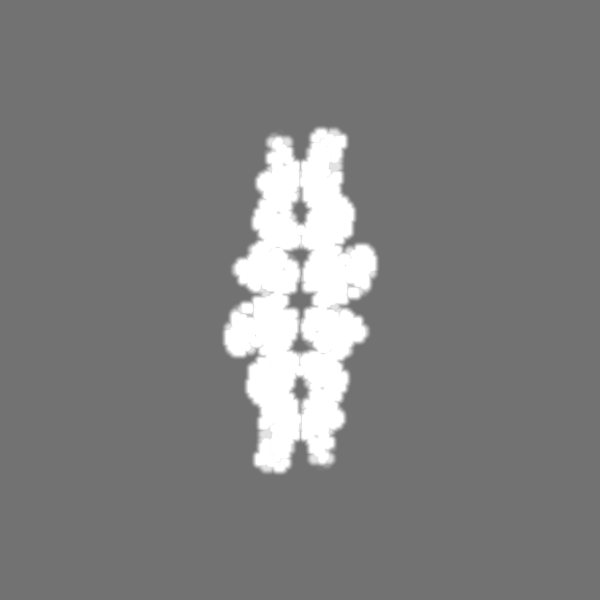
 Map data
 Sample
 Keywords
 Function and homology information
 Homo sapiens (human) /
 Authors
 United States, 2 items
  Citation
 Structure visualization
 Downloads & links
 emd_28025.png
 http://ftp.pdbj.org/pub/emdb/structures/EMD-28025

 Z (Sec.)
 Y (Row.)
 X (Col.)


 Sample components

 Processing
 Electron microscopy
 FIELD EMISSION GUN