Movie
Movie Controller

[English] 日本語
 Yorodumi
- EMDB-27960: D-cycloserine and glutamate bound Human GluN1a-GluN2C NMDA recept... -
+ Open data

- Basic information
| Entry |  | |||||||||
|---|---|---|---|---|---|---|---|---|---|---|
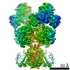
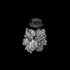
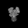
| Title | D-cycloserine and glutamate bound Human GluN1a-GluN2C NMDA receptor in nanodisc - splayed conformation | |||||||||
 Map data Map data | B-factor sharpened map | |||||||||
 Sample Sample |
| |||||||||
| Biological species |  Homo sapiens (human) Homo sapiens (human) | |||||||||
| Method | single particle reconstruction / cryo EM / Resolution: 3.28 Å | |||||||||
 Authors Authors | Chou T-H / Furukawa H | |||||||||
| Funding support |  United States, 2 items United States, 2 items
| |||||||||
 Citation Citation | Journal: Mol Cell / Year: 2022 Title: Structural insights into assembly and function of GluN1-2C, GluN1-2A-2C, and GluN1-2D NMDARs. Authors: Tsung-Han Chou / Hyunook Kang / Noriko Simorowski / Stephen F Traynelis / Hiro Furukawa / Abstract: Neurotransmission mediated by diverse subtypes of N-methyl-D-aspartate receptors (NMDARs) is fundamental for basic brain functions and development as well as neuropsychiatric diseases and disorders. ...Neurotransmission mediated by diverse subtypes of N-methyl-D-aspartate receptors (NMDARs) is fundamental for basic brain functions and development as well as neuropsychiatric diseases and disorders. NMDARs are glycine- and glutamate-gated ion channels that exist as heterotetramers composed of obligatory GluN1 and GluN2(A-D) and/or GluN3(A-B). The GluN2C and GluN2D subunits form ion channels with distinct properties and spatio-temporal expression patterns. Here, we provide the structures of the agonist-bound human GluN1-2C NMDAR in the presence and absence of the GluN2C-selective positive allosteric potentiator (PAM), PYD-106, the agonist-bound GluN1-2A-2C tri-heteromeric NMDAR, and agonist-bound GluN1-2D NMDARs by single-particle electron cryomicroscopy. Our analysis shows unique inter-subunit and domain arrangements of the GluN2C NMDARs, which contribute to functional regulation and formation of the PAM binding pocket and is distinct from GluN2D NMDARs. Our findings here provide the fundamental blueprint to study GluN2C- and GluN2D-containing NMDARs, which are uniquely involved in neuropsychiatric disorders. | |||||||||
| History |
|
- Structure visualization
| Supplemental images |
|---|

- Downloads & links
-EMDB archive
| Map data | emd_27960.map.gz | 229.5 MB |  EMDB map data format EMDB map data format | |
|---|---|---|---|---|
| Header (meta data) | emd-27960-v30.xmlemd-27960.xml | 17.5 KB 17.5 KB | Display Display | EMDB header |
| Images |  emd_27960.png emd_27960.png | 31.5 KB | ||
| Others | emd_27960_half_map_1.map.gzemd_27960_half_map_2.map.gz | 226 MB 226 MB | ||
| Archive directory |  http://ftp.pdbj.org/pub/emdb/structures/EMD-27960ftp://ftp.pdbj.org/pub/emdb/structures/EMD-27960 http://ftp.pdbj.org/pub/emdb/structures/EMD-27960ftp://ftp.pdbj.org/pub/emdb/structures/EMD-27960 | HTTPS FTP |
-Related structure data


-Links
| EMDB pages | EMDB (EBI/PDBe) / EMDataResource |
|---|
-Map
| File | Download / File: emd_27960.map.gz / Format: CCP4 / Size: 244.1 MB / Type: IMAGE STORED AS FLOATING POINT NUMBER (4 BYTES) | ||||||||||||||||||||||||||||||||||||
|---|---|---|---|---|---|---|---|---|---|---|---|---|---|---|---|---|---|---|---|---|---|---|---|---|---|---|---|---|---|---|---|---|---|---|---|---|---|
| Annotation | B-factor sharpened map | ||||||||||||||||||||||||||||||||||||
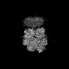
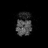
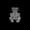
| Projections & slices | Image control
Images are generated by Spider. | ||||||||||||||||||||||||||||||||||||
| Voxel size | X=Y=Z: 0.856 Å | ||||||||||||||||||||||||||||||||||||
| Density |
| ||||||||||||||||||||||||||||||||||||
| Symmetry | Space group: 1 | ||||||||||||||||||||||||||||||||||||
| Details | EMDB XML:
|
 Z (Sec.)
 Y (Row.)
 X (Col.)


-Supplemental data
-Half map: Half map 1
| File | emd_27960_half_map_1.map | ||||||||||||
|---|---|---|---|---|---|---|---|---|---|---|---|---|---|
| Annotation | Half map 1 | ||||||||||||
| Projections & Slices |
| ||||||||||||
| Density Histograms |


-Half map: Half map 2
| File | emd_27960_half_map_2.map | ||||||||||||
|---|---|---|---|---|---|---|---|---|---|---|---|---|---|
| Annotation | Half map 2 | ||||||||||||
| Projections & Slices |
| ||||||||||||
| Density Histograms |


- Sample components
-Entire : Hetero-tetrameric GluN1a-GluN2C NMDA receptors
| Entire | Name: Hetero-tetrameric GluN1a-GluN2C NMDA receptors |
|---|---|
| Components |
|
-Supramolecule #1: Hetero-tetrameric GluN1a-GluN2C NMDA receptors
| Supramolecule | Name: Hetero-tetrameric GluN1a-GluN2C NMDA receptors / type: complex / Chimera: Yes / ID: 1 / Parent: 0 / Macromolecule list: all |
|---|---|
| Source (natural) | Organism: Homo sapiens (human) |
-Macromolecule #1: Ionotropic glutamate receptor, NMDA receptor GluN1a
| Macromolecule | Name: Ionotropic glutamate receptor, NMDA receptor GluN1a / type: protein_or_peptide / ID: 1 / Enantiomer: LEVO |
|---|---|
| Source (natural) | Organism: Homo sapiens (human) |
| Recombinant expression | Organism:   Spodoptera frugiperda (fall armyworm) Spodoptera frugiperda (fall armyworm) |
| Sequence | String: MSTMHLLTFA LLFSCSFARA ASDPKIVNIG AVLSTRKHEQ MFREAVNQAN KRHGSWKIQL NATSVTHKPN AIQMALSVCE DLISSQVYAI LVSHPPTPND HFTPTPVSYT AGFYRIPVLG LTTRMSIYSD KSIHLSFLRT VPPYSHQSSV WFEMMRVYSW NHIILLVSDD ...String: MSTMHLLTFA LLFSCSFARA ASDPKIVNIG AVLSTRKHEQ MFREAVNQAN KRHGSWKIQL NATSVTHKPN AIQMALSVCE DLISSQVYAI LVSHPPTPND HFTPTPVSYT AGFYRIPVLG LTTRMSIYSD KSIHLSFLRT VPPYSHQSSV WFEMMRVYSW NHIILLVSDD HEGRAAQKRL ETLLEERESK AEKVLQFDPG TKNVTALLME AKELEARVII LSASEDDAAT VYRAAAMLNM TGSGYVWLVG EREISGNALR YAPDGILGLQ LINGKNESAH ISDAVGVVAQ AVHELLEKEN ITDPPRGCVG NTNIWKTGPL FKRVLMSSKY ADGVTGRVEF NEDGDRKFAN YSIMNLQNRK LVQVGIYNGT HVIPNDRKII WPGGETEKPR GYQMSTRLKI VTIHQEPFVY VKPTLSDGTC KEEFTVNGDP VKKVICTGPN DTSPGSPRHT VPQCCYGFCI DLLIKLARTM NFTYEVHLVA DGKFGTQERV NNSNKKEWNG MMGELLSGQA DMIVAPLTIN NERAQYIEFS KPFKYQGLTI LVKKEIPRST LDSFMQPFQS TLWLLVGLSV HVVAVMLYLL DRFSPFGRFK VNSEEEEEDA LTLSSAMWFS WGVLLNSGIG EGAPRSFSAR ILGMVWAGFA MIIVASYTAN LAAFLVLDRP EERITGINDP RLRNPSDKFI YATVKQSSVD IYFRRQVELS TMYRHMEKHN YESAAEAIQA VRDNKLHAFI WDSAVLEFEA SQKCDLVTTG ELFFRSGFGI GMRKDSPWKQ NVSLSILKSH ENGFMEDLDK TWVRYQECDS RSNAPATLTF ENMAGVFMLV AGGIVAGIFL IFIEIAYKRH KDANGAQ |
-Macromolecule #2: Ionotropic glutamate receptor, NMDA receptor GluN2C
| Macromolecule | Name: Ionotropic glutamate receptor, NMDA receptor GluN2C / type: protein_or_peptide / ID: 2 / Enantiomer: LEVO |
|---|---|
| Source (natural) | Organism: Homo sapiens (human) |
| Recombinant expression | Organism: Spodoptera frugiperda (fall armyworm) |
| Sequence | String: MGTMRLFLLA VLFLFSFARA TGWSHPQFEK GGGSGGGSGG SAWSHPQFEK GALVPRGEQG MTVAVVFSSS GPPQAQFRAR LTPQSFLDLP LEIQPLTVGV NTTNPSSLLT QICGLLGAAH VHGIVFEDNV DTEAVAQILD FISSQTHVPI LSISGGSAVV LTPKEPGSAF ...String: MGTMRLFLLA VLFLFSFARA TGWSHPQFEK GGGSGGGSGG SAWSHPQFEK GALVPRGEQG MTVAVVFSSS GPPQAQFRAR LTPQSFLDLP LEIQPLTVGV NTTNPSSLLT QICGLLGAAH VHGIVFEDNV DTEAVAQILD FISSQTHVPI LSISGGSAVV LTPKEPGSAF LQLGVSLEQQ LQVLFKVLEE YDWSAFAVIT SLHPGHALFL EGVRAVADAS HVSWRLLDVV TLELGPGGPR ARTQRLLRQL DAPVFVAYCS REEAEVLFAE AAQAGLVGPG HVWLVPNLAL GSTDAPPATF PVGLISVVTE SWRLSLRQKV RDGVAILALG AHSYWRQHGT LPAPAGDCRV HPGPVSPARE AFYRHLLNVT WEGRDFSFSP GGYLVQPTMV VIALNRHRLW EMVGRWEHGV LYMKYPVWPR YSASLQPVVD SRHLTVATLE ERPFVIVESP DPGTGGCVPN TVPCRRQSNH TFSSGDVAPY TKLCCKGFCI DILKKLARVV KFSYDLYLVT NGKHGKRVRG VWNGMIGEVY YKRADMAIGS LTINEERSEI VDFSVPFVET GISVMVARSN GTVSPSAFLE PYSPAVWVMM FVMCLTVVAI TVFMFEYFSP VSYNQNLTRG KKSGGPAFTI GKSVWLLWAL VFNNSVPIEN PRGTTSKIMV LVWAFFAVIF LASYTANLAA FMIQEQYIDT VSGLSDKKFQ RPQDQYPPFR FGTVPNGSTE RNIRSNYRDM HTHMVKFNQR SVEDALTSLK MGKLDAFIYD AAVLNYMAGK DEGCKLVTIG SGKVFATTGY GIAMQKDSHW KRAIDLALLQ FLGDGETQKL ETVWLSGICQ NEKNEVMSSK LDIDNMAGVF YMLLVAMGLA LLVFAWEHLV YWKLRHSVPN |
-Experimental details
-Structure determination
| Method | cryo EM |
|---|---|
 Processing Processing | single particle reconstruction |
| Aggregation state | particle |
-Sample preparation
| Concentration | 4 mg/mL |
|---|---|
| Buffer | pH: 7.5 |
| Vitrification | Cryogen name: ETHANE / Chamber humidity: 85 % / Chamber temperature: 285 K / Instrument: FEI VITROBOT MARK IV |
- Electron microscopy
| Microscope | FEI TITAN KRIOS |
|---|---|
| Image recording | Film or detector model: GATAN K3 BIOQUANTUM (6k x 4k) / Average electron dose: 57.6 e/Å2 |
| Electron beam | Acceleration voltage: 300 kV / Electron source:  FIELD EMISSION GUN FIELD EMISSION GUN |
| Electron optics | Illumination mode: FLOOD BEAM / Imaging mode: BRIGHT FIELD / Nominal defocus max: 2.8000000000000003 µm / Nominal defocus min: 1.4000000000000001 µm |
| Experimental equipment |  Model: Titan Krios / Image courtesy: FEI Company |
-Image processing
| Startup model | Type of model: EMDB MAP EMDB ID: |
|---|---|
| Final reconstruction | Resolution.type: BY AUTHOR / Resolution: 3.28 Å / Resolution method: FSC 0.143 CUT-OFF / Software - Name: cryoSPARC (ver. 3.3.2) / Number images used: 111641 |
| Initial angle assignment | Type: MAXIMUM LIKELIHOOD |
| Final angle assignment | Type: MAXIMUM LIKELIHOOD |