 Movie
Movie Controller


+ Open data

- Basic information
| Entry |  | |||||||||
|---|---|---|---|---|---|---|---|---|---|---|
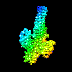
| Title | Cryo-EM structure of proximal fiber in del7.3K2R1 free phage | |||||||||
 Map data Map data | ||||||||||
 Sample Sample |
| |||||||||
 Keywords Keywords | Bacteriophage T7 / VIRUS | |||||||||
| Function / homology |  Function and homology information Function and homology informationvirus tail, fiber / adhesion receptor-mediated virion attachment to host cell / symbiont entry into host cell / virion attachment to host cell / identical protein binding Similarity search - Function | |||||||||
| Biological species |   Escherichia phage T7 (virus) Escherichia phage T7 (virus) | |||||||||
| Method | single particle reconstruction / cryo EM / Resolution: 3.3 Å | |||||||||
 Authors Authors | Wang C / Liu J / Molineux IJ | |||||||||
| Funding support |  United States, 1 items United States, 1 items
| |||||||||
 Citation Citation | Journal: To Be Published Title: Asymmetric reconstruction of gp8-gp11 with terminal dsDNA associated from the del7.3K2R1. Authors: Wang C / Liu J / Molineux IJ | |||||||||
| History |
|
- Structure visualization
| Supplemental images |
|---|
- Downloads & links
-EMDB archive
| Map data | emd_27657.map.gz | 2.9 MB | EMDB map data format | |
|---|---|---|---|---|
| Header (meta data) | emd-27657-v30.xmlemd-27657.xml | 13.1 KB 13.1 KB | Display Display | EMDB header |
| Images |  emd_27657.png emd_27657.png | 40.3 KB | ||
| Filedesc metadata | emd-27657.cif.gz | 3.7 KB | ||
| Others | emd_27657_additional_1.map.gzemd_27657_half_map_1.map.gzemd_27657_half_map_2.map.gz | 191.1 MB 191.9 MB 191.9 MB | ||
| Archive directory |  http://ftp.pdbj.org/pub/emdb/structures/EMD-27657ftp://ftp.pdbj.org/pub/emdb/structures/EMD-27657 http://ftp.pdbj.org/pub/emdb/structures/EMD-27657ftp://ftp.pdbj.org/pub/emdb/structures/EMD-27657 | HTTPS FTP |
-Related structure data
| Related structure data |  8dspMC M: atomic model generated by this map C: citing same article ( |
|---|---|
| Similar structure data |

-Links
| EMDB pages | EMDB (EBI/PDBe) / EMDataResource |
|---|
-Map
| File | Download / File: emd_27657.map.gz / Format: CCP4 / Size: 244.1 MB / Type: IMAGE STORED AS FLOATING POINT NUMBER (4 BYTES) | ||||||||||||||||||||||||||||||||||||
|---|---|---|---|---|---|---|---|---|---|---|---|---|---|---|---|---|---|---|---|---|---|---|---|---|---|---|---|---|---|---|---|---|---|---|---|---|---|
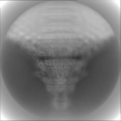
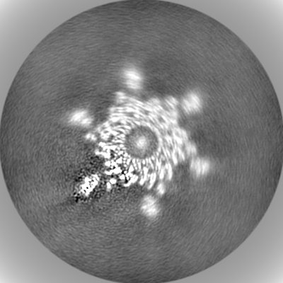
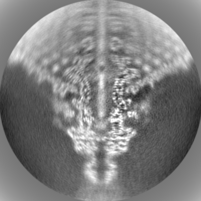
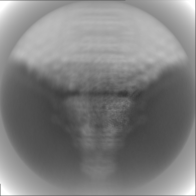
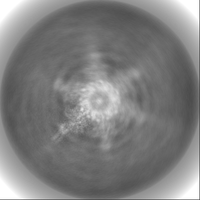
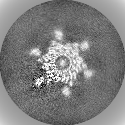
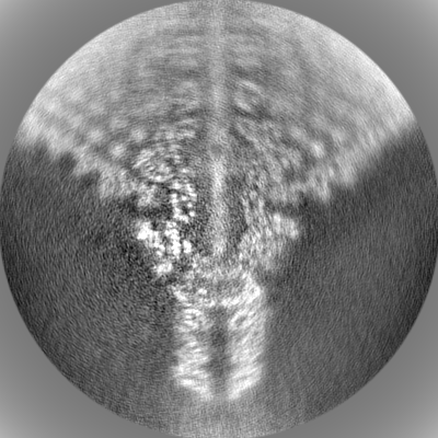
| Projections & slices | Image control
Images are generated by Spider. | ||||||||||||||||||||||||||||||||||||
| Voxel size | X=Y=Z: 1.068 Å | ||||||||||||||||||||||||||||||||||||
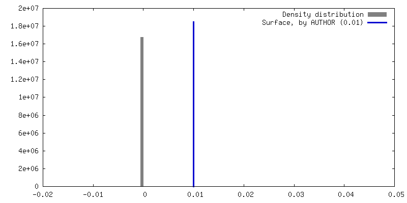
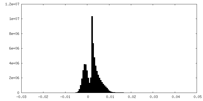
| Density |
| ||||||||||||||||||||||||||||||||||||
| Symmetry | Space group: 1 | ||||||||||||||||||||||||||||||||||||
| Details | EMDB XML:
|
 Z (Sec.)
 Y (Row.)
 X (Col.)


-Supplemental data
-Additional map: unmasked average
| File | emd_27657_additional_1.map | ||||||||||||
|---|---|---|---|---|---|---|---|---|---|---|---|---|---|
| Annotation | unmasked average | ||||||||||||
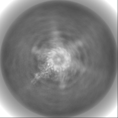
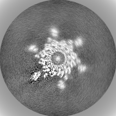
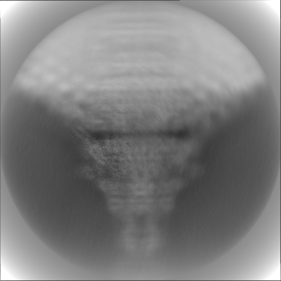
| Projections & Slices |
| ||||||||||||
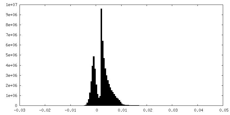
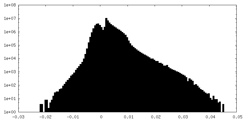
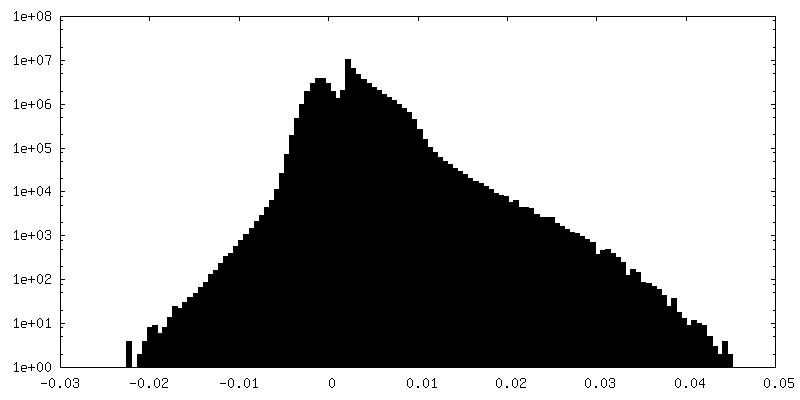
| Density Histograms |


-Half map: #1
| File | emd_27657_half_map_1.map | ||||||||||||
|---|---|---|---|---|---|---|---|---|---|---|---|---|---|
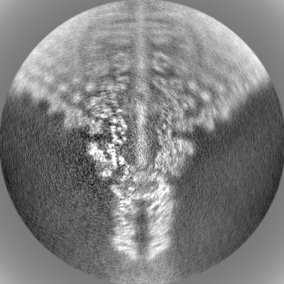
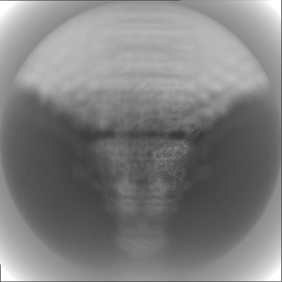
| Projections & Slices |
| ||||||||||||
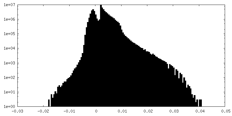
| Density Histograms |


-Half map: #2
| File | emd_27657_half_map_2.map | ||||||||||||
|---|---|---|---|---|---|---|---|---|---|---|---|---|---|
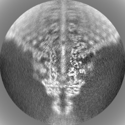
| Projections & Slices |
| ||||||||||||
| Density Histograms |

- Sample components
-Entire : Escherichia phage T7
| Entire | Name: Escherichia phage T7 (virus) |
|---|---|
| Components |
|
-Supramolecule #1: Escherichia phage T7
| Supramolecule | Name: Escherichia phage T7 / type: virus / ID: 1 / Parent: 0 / NCBI-ID: 10760 / Sci species name: Escherichia phage T7 / Virus type: VIRION / Virus isolate: STRAIN / Virus enveloped: Yes / Virus empty: No |
|---|
-Experimental details
-Structure determination
| Method | cryo EM |
|---|---|
 Processing Processing | single particle reconstruction |
| Aggregation state | particle |
-Sample preparation
| Buffer | pH: 7.5 |
|---|---|
| Vitrification | Cryogen name: ETHANE |
- Electron microscopy
| Microscope | FEI TITAN KRIOS |
|---|---|
| Image recording | Film or detector model: GATAN K3 (6k x 4k) / Average electron dose: 30.0 e/Å2 |
| Electron beam | Acceleration voltage: 300 kV / Electron source:  FIELD EMISSION GUN FIELD EMISSION GUN |
| Electron optics | Illumination mode: FLOOD BEAM / Imaging mode: BRIGHT FIELD / Nominal defocus max: 1.2 µm / Nominal defocus min: 0.6 µm |
| Experimental equipment |  Model: Titan Krios / Image courtesy: FEI Company |
-Image processing
| Startup model | Type of model: OTHER |
|---|---|
| Final reconstruction | Resolution.type: BY AUTHOR / Resolution: 3.3 Å / Resolution method: FSC 0.143 CUT-OFF / Number images used: 99318 |
| Initial angle assignment | Type: MAXIMUM LIKELIHOOD |
| Final angle assignment | Type: MAXIMUM LIKELIHOOD |
