National Institutes of Health/National Institute Of Allergy and Infectious Diseases (NIH/NIAID)
AI151055
米国
National Institutes of Health/National Institute of General Medical Sciences (NIH/NIGMS)
GM071940
米国
引用
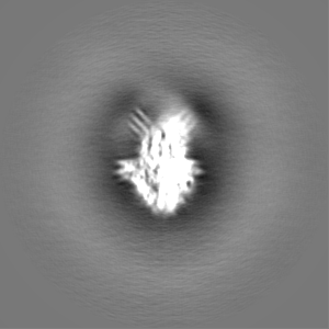
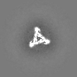
ジャーナル: mBio / 年: 2022 タイトル: Atomic Structure of IglD Demonstrates Its Role as a Component of the Baseplate Complex of the Type VI Secretion System. 著者: Xiaoyu Liu / Daniel L Clemens / Bai-Yu Lee / Xue Yang / Z Hong Zhou / Marcus A Horwitz / 要旨: Francisella tularensis, a Tier 1 select agent of bioterrorism, contains a type VI secretion system (T6SS) encoded within the pathogenicity island (FPI), which is critical for its pathogenesis. Among ...Francisella tularensis, a Tier 1 select agent of bioterrorism, contains a type VI secretion system (T6SS) encoded within the pathogenicity island (FPI), which is critical for its pathogenesis. Among the 18 proteins encoded by FPI is IglD, which is essential to 's intracellular growth and virulence, but neither its location within T6SS nor its functional role has been established. Here, we present the cryoEM structure of IglD from Francisella novicida and show that the IglD forms a homotrimer that is structurally homologous to the T6SS baseplate protein TssK in Escherichia coli. Each IglD monomer consists of an N-terminal β-sandwich domain, a 4-helix bundle domain, and a flexible C-terminal domain. While the overall folds of IglD and TssK are similar, the two structures differ in three aspects: the relative orientation between their β-sandwich and the 4-helix bundle domains; two insertion loops present in TssK's β-sandwich domain; and, consequently, a lack of subunit-subunit interaction between insertion loops in the IglD trimer. Phylogenetic analysis indicates that IglD is genetically remote from the TssK orthologs in other T6SSs. While the other components of the baseplate are unknown, we conducted pulldown assays showing IglJ interacts with IglD and IglH, pointing to a model wherein IglD, IglH, and IglJ form the baseplate of the T6SS. Alanine substitution mutagenesis further established that IglD's hydrophobic pocket in the N-terminal β-sandwich domain interacts with two loops of IglJ, reminiscent of the TssK-TssG interaction. These results form a framework for understanding the hitherto unexplored T6SS baseplate. Francisella tularensis is a facultatively intracellular Gram-negative bacterium that causes the serious and potentially fatal zoonotic illness, tularemia. Because of its extraordinarily high infectivity and mortality to humans, especially when inhaled, F. tularensis is considered a potential bioterrorism agent and is classified as a Tier 1 select agent. The type VI secretion system (T6SS) encoded within the pathogenicity island (FPI) is critical to its pathogenesis, but its baseplate components are largely unknown. Here, we report the cryoEM structure of IglD from Francisella novicida and demonstrate its role as a component of the baseplate complex of the T6SS. We further show that IglD interacts with IglJ and IglH, and propose a model in which these proteins interact to form the T6SS baseplate. Elucidation of the structure and composition of the baseplate should facilitate the design of strategies to prevent and treat infections caused by F. tularensis.
 ムービー
ムービー コントローラー


 データを開く
 基本情報
基本情報
 マップデータ
 試料
試料 キーワード
キーワード 機能・相同性情報
 Francisella tularensis subsp. novicida (バクテリア)
 データ登録者
 米国, 2件
  引用
引用
 構造の表示
構造の表示
 ダウンロードとリンク
 emd_27656.png
 http://ftp.pdbj.org/pub/emdb/structures/EMD-27656
 Z (Sec.)
 Y (Row.)
 X (Col.)


 試料の構成要素
 解析
解析 電子顕微鏡法
 FIELD EMISSION GUN
