 Movie
Movie Controller

[English] 日本語
 Yorodumi
- EMDB-27553: Cryo-EM structure of human Glycine Receptor alpha1-beta heteromer... -
+ Open data

- Basic information
| Entry |  | |||||||||
|---|---|---|---|---|---|---|---|---|---|---|
| Title | Cryo-EM structure of human Glycine Receptor alpha1-beta heteromer, apo state | |||||||||
 Map data Map data | ||||||||||
 Sample Sample |
| |||||||||
 Keywords Keywords | glycine receptor subunit alpha-1 / glycine receptor subunit beta / Green fluorescent protein / MEMBRANE PROTEIN | |||||||||
| Function / homology |  Function and homology information Function and homology informationtaurine binding / positive regulation of acrosome reaction / negative regulation of transmission of nerve impulse / glycine-gated chloride channel complex / synaptic transmission, glycinergic / Neurotransmitter receptors and postsynaptic signal transmission / postsynaptic specialization / extracellularly glycine-gated ion channel activity / extracellularly glycine-gated chloride channel activity / inhibitory synapse ...taurine binding / positive regulation of acrosome reaction / negative regulation of transmission of nerve impulse / glycine-gated chloride channel complex / synaptic transmission, glycinergic / Neurotransmitter receptors and postsynaptic signal transmission / postsynaptic specialization / extracellularly glycine-gated ion channel activity / extracellularly glycine-gated chloride channel activity / inhibitory synapse / glycinergic synapse / response to alcohol / excitatory extracellular ligand-gated monoatomic ion channel activity / chloride transport / cellular response to zinc ion / inhibitory postsynaptic potential / startle response / glycine binding / intracellular membrane-bounded organelle / cellular response to ethanol / chloride channel complex / monoatomic ion transport / muscle contraction / ligand-gated monoatomic ion channel activity involved in regulation of presynaptic membrane potential / chloride transmembrane transport / bioluminescence / cellular response to amino acid stimulus / generation of precursor metabolites and energy / neuropeptide signaling pathway / transmitter-gated monoatomic ion channel activity involved in regulation of postsynaptic membrane potential / regulation of membrane potential / GABA-ergic synapse / transmembrane signaling receptor activity / nervous system development / chemical synaptic transmission / perikaryon / postsynaptic membrane / neuron projection / external side of plasma membrane / neuronal cell body / synapse / dendrite / protein-containing complex binding / zinc ion binding / membrane / identical protein binding / plasma membrane / cytoplasm Similarity search - Function | |||||||||
| Biological species |  Homo sapiens (human) Homo sapiens (human) | |||||||||
| Method | single particle reconstruction / cryo EM / Resolution: 3.55 Å | |||||||||
 Authors Authors | Liu X / Wang W | |||||||||
| Funding support |  United States, 1 items United States, 1 items
| |||||||||
 Citation Citation | Journal: Nat Commun / Year: 2023 Title: Asymmetric gating of a human hetero-pentameric glycine receptor. Authors: Xiaofen Liu / Weiwei Wang / Abstract: Hetero-pentameric Cys-loop receptors constitute a major type of neurotransmitter receptors that enable signal transmission and processing in the nervous system. Despite intense investigations into ...Hetero-pentameric Cys-loop receptors constitute a major type of neurotransmitter receptors that enable signal transmission and processing in the nervous system. Despite intense investigations into their working mechanism and pharmaceutical potentials, how neurotransmitters activate these receptors remains unclear due to the lack of high-resolution structural information in the activated open state. Here we report near-atomic resolution structures resolved in digitonin consistent with all principle functional states of the human α1β GlyR, which is a major Cys-loop receptor that mediates inhibitory neurotransmission in the central nervous system of adults. Glycine binding induces cooperative and symmetric structural rearrangements in the neurotransmitter-binding extracellular domain but asymmetrical pore dilation in the transmembrane domain. Symmetric response in the extracellular domain is consistent with electrophysiological data showing cooperative glycine activation and contribution from both α1 and β subunits. A set of functionally essential but differentially charged amino acid residues in the transmembrane domain of the α1 and β subunits explains asymmetric activation. These findings provide a foundation for understanding how the gating of the Cys-loop receptor family members diverges to accommodate specific physiological environments. | |||||||||
| History |
|
- Structure visualization
| Supplemental images |
|---|

- Downloads & links
-EMDB archive
| Map data | emd_27553.map.gz | 86 MB | EMDB map data format | |
|---|---|---|---|---|
| Header (meta data) | emd-27553-v30.xmlemd-27553.xml | 20.2 KB 20.2 KB | Display Display | EMDB header |
| Images | 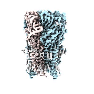 emd_27553.png emd_27553.png | 104.2 KB | ||
| Filedesc metadata | emd-27553.cif.gz | 7 KB | ||
| Others | emd_27553_half_map_1.map.gzemd_27553_half_map_2.map.gz | 84.7 MB 84.7 MB | ||
| Archive directory |  http://ftp.pdbj.org/pub/emdb/structures/EMD-27553ftp://ftp.pdbj.org/pub/emdb/structures/EMD-27553 http://ftp.pdbj.org/pub/emdb/structures/EMD-27553ftp://ftp.pdbj.org/pub/emdb/structures/EMD-27553 | HTTPS FTP |
-Related structure data
| Related structure data |  8dn3MC  8dn2C  8dn4C  8dn5C M: atomic model generated by this map C: citing same article ( |
|---|---|
| Similar structure data |


-Links
| EMDB pages | EMDB (EBI/PDBe) / EMDataResource |
|---|---|
| Related items in Molecule of the Month |


-Map
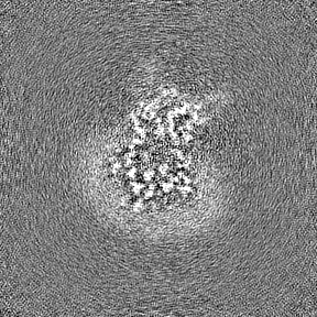
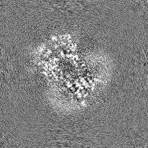
| File | Download / File: emd_27553.map.gz / Format: CCP4 / Size: 91.1 MB / Type: IMAGE STORED AS FLOATING POINT NUMBER (4 BYTES) | ||||||||||||||||||||||||||||||||||||
|---|---|---|---|---|---|---|---|---|---|---|---|---|---|---|---|---|---|---|---|---|---|---|---|---|---|---|---|---|---|---|---|---|---|---|---|---|---|
| Projections & slices | Image control
Images are generated by Spider. | ||||||||||||||||||||||||||||||||||||
| Voxel size | X=Y=Z: 0.83 Å | ||||||||||||||||||||||||||||||||||||
| Density |
| ||||||||||||||||||||||||||||||||||||
| Symmetry | Space group: 1 | ||||||||||||||||||||||||||||||||||||
| Details | EMDB XML:
|
 Z (Sec.)
 Y (Row.)
 X (Col.)


-Supplemental data
-Half map: #2
| File | emd_27553_half_map_1.map | ||||||||||||
|---|---|---|---|---|---|---|---|---|---|---|---|---|---|
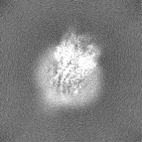
| Projections & Slices |
| ||||||||||||
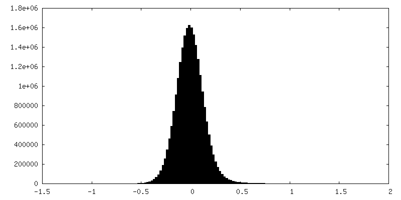
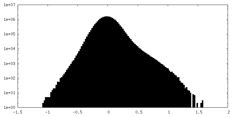
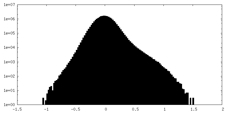
| Density Histograms |


-Half map: #1
| File | emd_27553_half_map_2.map | ||||||||||||
|---|---|---|---|---|---|---|---|---|---|---|---|---|---|
| Projections & Slices |
| ||||||||||||
| Density Histograms |

- Sample components
+Entire : heteromeric glycine receptor alpha-1 and beta, apo state
+Supramolecule #1: heteromeric glycine receptor alpha-1 and beta, apo state
+Macromolecule #1: Glycine receptor subunit alpha-1
+Macromolecule #2: Glycine receptor subunit beta,Green fluorescent protein,Glycine r...
+Macromolecule #3: 2-acetamido-2-deoxy-beta-D-glucopyranose

+Macromolecule #4: HEXANE
+Macromolecule #5: UNDECANE
+Macromolecule #6: nonane
+Macromolecule #7: N-BUTANE

+Macromolecule #8: HEPTANE
+Macromolecule #9: DECANE

+Macromolecule #10: CHLORIDE ION
-Experimental details
-Structure determination
| Method | cryo EM |
|---|---|
 Processing Processing | single particle reconstruction |
| Aggregation state | particle |
-Sample preparation
| Concentration | 6 mg/mL | ||||||||||||
|---|---|---|---|---|---|---|---|---|---|---|---|---|---|
| Buffer | pH: 8 Component:
| ||||||||||||
| Grid | Model: Quantifoil R1.2/1.3 / Material: GOLD / Mesh: 400 / Support film - Material: GOLD / Support film - topology: HOLEY / Pretreatment - Type: GLOW DISCHARGE / Pretreatment - Time: 70 sec. | ||||||||||||
| Vitrification | Cryogen name: ETHANE / Chamber humidity: 100 % |
- Electron microscopy
| Microscope | FEI TITAN KRIOS |
|---|---|
| Image recording | Film or detector model: GATAN K3 (6k x 4k) / Average electron dose: 69.6 e/Å2 |
| Electron beam | Acceleration voltage: 300 kV / Electron source:  FIELD EMISSION GUN FIELD EMISSION GUN |
| Electron optics | Illumination mode: FLOOD BEAM / Imaging mode: BRIGHT FIELD / Nominal defocus max: 2.5 µm / Nominal defocus min: 1.0 µm |
| Sample stage | Specimen holder model: GATAN LIQUID NITROGEN / Cooling holder cryogen: NITROGEN |
| Experimental equipment |  Model: Titan Krios / Image courtesy: FEI Company |
-Image processing
| Startup model | Type of model: PDB ENTRY PDB model - PDB ID: |
|---|---|
| Final reconstruction | Applied symmetry - Point group: C1 (asymmetric) / Resolution.type: BY AUTHOR / Resolution: 3.55 Å / Resolution method: FSC 0.143 CUT-OFF / Number images used: 29850 |
| Initial angle assignment | Type: MAXIMUM LIKELIHOOD |
| Final angle assignment | Type: MAXIMUM LIKELIHOOD |

