 ムービー
ムービー コントローラー


+ データを開く

- 基本情報
基本情報
| 登録情報 | データベース: EMDB / ID: EMD-2754 | |||||||||
|---|---|---|---|---|---|---|---|---|---|---|
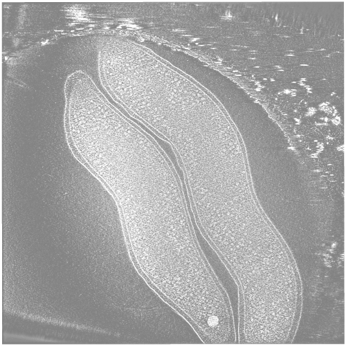
| タイトル | Electron cryo-tomography of Campylobacter jejuni | |||||||||
 マップデータ マップデータ | Electron cryo-tomogram of two Campylobacter jejuni cells | |||||||||
 試料 試料 |
| |||||||||
 キーワード キーワード | food poisoning / cryo electron tomography / cryo-EM / chemoreceptors / acidocalcisomes | |||||||||
| 生物種 |   Campylobacter jejuni (カンピロバクター) Campylobacter jejuni (カンピロバクター) | |||||||||
| 手法 | 電子線トモグラフィー法 / クライオ電子顕微鏡法 | |||||||||
 データ登録者 データ登録者 | Muller A / Beeby M / McDowall AW / Chow J / Jensen GJ / Clemons WM | |||||||||
 引用 引用 | ジャーナル: Microbiologyopen / 年: 2014 タイトル: Ultrastructure and complex polar architecture of the human pathogen Campylobacter jejuni. 著者: Axel Müller / Morgan Beeby / Alasdair W McDowall / Janet Chow / Grant J Jensen / William M Clemons /  要旨: Campylobacter jejuni is one of the most successful food-borne human pathogens. Here we use electron cryotomography to explore the ultrastructure of C. jejuni cells in logarithmically growing cultures. ...Campylobacter jejuni is one of the most successful food-borne human pathogens. Here we use electron cryotomography to explore the ultrastructure of C. jejuni cells in logarithmically growing cultures. This provides the first look at this pathogen in a near-native state at macromolecular resolution (~5 nm). We find a surprisingly complex polar architecture that includes ribosome exclusion zones, polyphosphate storage granules, extensive collar-shaped chemoreceptor arrays, and elaborate flagellar motors. | |||||||||
| 履歴 |
|
- 構造の表示
構造の表示
| ムービー |
 ムービービューア ムービービューア |
|---|---|
| 構造ビューア | EMマップ: SurfViewMolmilJmol/JSmol |
| 添付画像 |
- ダウンロードとリンク
-EMDBアーカイブ
| マップデータ | emd_2754.map.gz | 157.8 MB | EMDBマップデータ形式 | |
|---|---|---|---|---|
| ヘッダ (付随情報) | emd-2754-v30.xmlemd-2754.xml | 10.2 KB 10.2 KB | 表示 表示 | EMDBヘッダ |
| 画像 |  EMD-2754.png EMD-2754.png | 286 KB | ||
| アーカイブディレクトリ |  http://ftp.pdbj.org/pub/emdb/structures/EMD-2754ftp://ftp.pdbj.org/pub/emdb/structures/EMD-2754 http://ftp.pdbj.org/pub/emdb/structures/EMD-2754ftp://ftp.pdbj.org/pub/emdb/structures/EMD-2754 | HTTPS FTP |
-検証レポート
| 文書・要旨 | emd_2754_validation.pdf.gz | 141.8 KB | 表示 | EMDB検証レポート |
|---|---|---|---|---|
| 文書・詳細版 | emd_2754_full_validation.pdf.gz | 140.7 KB | 表示 | |
| XML形式データ | emd_2754_validation.xml.gz | 4.3 KB | 表示 | |
| アーカイブディレクトリ | https://ftp.pdbj.org/pub/emdb/validation_reports/EMD-2754ftp://ftp.pdbj.org/pub/emdb/validation_reports/EMD-2754 | HTTPS FTP |
-リンク
| EMDBのページ | EMDB (EBI/PDBe) / EMDataResource |
|---|
-マップ
| ファイル | ダウンロード / ファイル: emd_2754.map.gz / 形式: CCP4 / 大きさ: 390.6 MB / タイプ: IMAGE STORED AS SIGNED BYTE | ||||||||||||||||||||||||||||||||||||||||||||||||||||||||||||||||||||
|---|---|---|---|---|---|---|---|---|---|---|---|---|---|---|---|---|---|---|---|---|---|---|---|---|---|---|---|---|---|---|---|---|---|---|---|---|---|---|---|---|---|---|---|---|---|---|---|---|---|---|---|---|---|---|---|---|---|---|---|---|---|---|---|---|---|---|---|---|---|
| 注釈 | Electron cryo-tomogram of two Campylobacter jejuni cells | ||||||||||||||||||||||||||||||||||||||||||||||||||||||||||||||||||||
| ボクセルのサイズ | X=Y=Z: 19.24 Å | ||||||||||||||||||||||||||||||||||||||||||||||||||||||||||||||||||||
| 密度 |
| ||||||||||||||||||||||||||||||||||||||||||||||||||||||||||||||||||||
| 対称性 | 空間群: 1 | ||||||||||||||||||||||||||||||||||||||||||||||||||||||||||||||||||||
| 詳細 | EMDB XML:
CCP4マップ ヘッダ情報:
| ||||||||||||||||||||||||||||||||||||||||||||||||||||||||||||||||||||
-添付データ
- 試料の構成要素
-全体 : Campylobacter jejuni subsp. jejuni ATCC 29428 (whole bacterial cells)
| 全体 | 名称: Campylobacter jejuni subsp. jejuni ATCC 29428 (whole bacterial cells) |
|---|---|
| 要素 |
|
-超分子 #1000: Campylobacter jejuni subsp. jejuni ATCC 29428 (whole bacterial cells)
| 超分子 | 名称: Campylobacter jejuni subsp. jejuni ATCC 29428 (whole bacterial cells) タイプ: sample / ID: 1000 / Number unique components: 1 |
|---|
-超分子 #1: Campylobacter jejuni
| 超分子 | 名称: Campylobacter jejuni / タイプ: organelle_or_cellular_component / ID: 1 / コピー数: 2 / 組換発現: No / データベース: NCBI |
|---|---|
| 由来(天然) | 生物種: Campylobacter jejuni (カンピロバクター) / 株: ATCC 29428 / 別称: Campylobacter jejuni |
-実験情報
-構造解析
| 手法 | クライオ電子顕微鏡法 |
|---|---|
 解析 解析 | 電子線トモグラフィー法 |
| 試料の集合状態 | cell |
-試料調製
| グリッド | 詳細: Quantifoil |
|---|---|
| 凍結 | 凍結剤: ETHANE-PROPANE MIXTURE / チャンバー内湿度: 100 % / チャンバー内温度: 77 K / 装置: FEI VITROBOT MARK III 手法: Sample preparation closely followed established procedures (Iancu et al., 2007). In brief: 4 microlitres of a 10 nm colloidal gold (Sigma, USA) in 5% BSA was added to 16 microlitres of a C. ...手法: Sample preparation closely followed established procedures (Iancu et al., 2007). In brief: 4 microlitres of a 10 nm colloidal gold (Sigma, USA) in 5% BSA was added to 16 microlitres of a C. jejuni culture that had been allowed to grow to an optical density of 0.5. 3 microlitres of this mix were then placed onto a glow discharged carbon-coated R 2/2 Quantifoil grid in a Vitrobot (FEI Company, Hillsboro, OR, USA). Prior to this a 10 nm colloidal gold suspension in 5% BSA solution was added to the Quantifoil grid and allowed to dry. The temperature in the Vitrobot chamber was kept at 22 degrees Celsius with 100% humidity. Placing the sample onto the grid was followed by a 1 second blot with an offset of -1.5 degrees, a drain time of 1 second, and plunge-frozen in a mixture of liquid ethane (63%) and propane (37%). The frozen grids were than stored in liquid nitrogen until further use. |
- 電子顕微鏡法
| 顕微鏡 | FEI POLARA 300 |
|---|---|
| 温度 | 最低: 76.9 K / 最高: 77.1 K / 平均: 77 K |
| 特殊光学系 | エネルギーフィルター - 名称: GATAN GIF エネルギーフィルター - エネルギー下限: 0.0 eV エネルギーフィルター - エネルギー上限: 20.0 eV |
| 詳細 | Camera post-energy filter |
| 日付 | 2014年1月1日 |
| 撮影 | 実像数: 121 / 平均電子線量: 200 e/Å2 / ビット/ピクセル: 16 |
| 電子線 | 加速電圧: 300 kV / 電子線源:  FIELD EMISSION GUN FIELD EMISSION GUN |
| 電子光学系 | 照射モード: OTHER / 撮影モード: BRIGHT FIELD / Cs: 2.2 mm / 最大 デフォーカス(公称値): -12.0 µm / 倍率(公称値): 22500 |
| 試料ステージ | 試料ホルダー: Liquid nitrogen cooled / 試料ホルダーモデル: GATAN HELIUM / Tilt series - Axis1 - Min angle: -60 ° / Tilt series - Axis1 - Max angle: 60 ° / Tilt series - Axis1 - Angle increment: 1 ° |
| 実験機器 |  モデル: Tecnai Polara / 画像提供: FEI Company |
-画像解析
| 詳細 | Single-axis tilt series from -60 degrees to 60 degrees with images were collected in 1 degree increments and an under-focus of 12 microns using a 300 keV FEI Polara FEG TEM controlled by Leginon software (Suloway et al., 2009) and the cumulative dose was not allowed to exceed 200 e A2. The images were recorded on a 4096 x 4096 pixel Ultracam (Gatan, Pleasanton, CA, USA) at a magnification of 22500 (0.98 nm/pixel). The IMOD software package (Kremer et al., 1996) was used to calculate 3D reconstructions. |
|---|---|
| 最終 再構成 | アルゴリズム: OTHER / ソフトウェア - 名称: IMOD, tomo3d 詳細: SIRT reconstruction of a fine-aligned stack low-pass filtered to first zero at approximately 5.5 nanometres. 使用した粒子像数: 121 |
