 Movie
Movie Controller


+ Open data

- Basic information
| Entry | Database: EMDB / ID: EMD-2754 | |||||||||
|---|---|---|---|---|---|---|---|---|---|---|
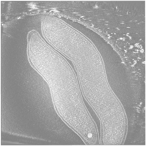
| Title | Electron cryo-tomography of Campylobacter jejuni | |||||||||
 Map data Map data | Electron cryo-tomogram of two Campylobacter jejuni cells | |||||||||
 Sample Sample |
| |||||||||
 Keywords Keywords | food poisoning / cryo electron tomography / cryo-EM / chemoreceptors / acidocalcisomes | |||||||||
| Biological species |   Campylobacter jejuni (Campylobacter) Campylobacter jejuni (Campylobacter) | |||||||||
| Method | electron tomography / cryo EM | |||||||||
 Authors Authors | Muller A / Beeby M / McDowall AW / Chow J / Jensen GJ / Clemons WM | |||||||||
 Citation Citation | Journal: Microbiologyopen / Year: 2014 Title: Ultrastructure and complex polar architecture of the human pathogen Campylobacter jejuni. Authors: Axel Müller / Morgan Beeby / Alasdair W McDowall / Janet Chow / Grant J Jensen / William M Clemons /  Abstract: Campylobacter jejuni is one of the most successful food-borne human pathogens. Here we use electron cryotomography to explore the ultrastructure of C. jejuni cells in logarithmically growing cultures. ...Campylobacter jejuni is one of the most successful food-borne human pathogens. Here we use electron cryotomography to explore the ultrastructure of C. jejuni cells in logarithmically growing cultures. This provides the first look at this pathogen in a near-native state at macromolecular resolution (~5 nm). We find a surprisingly complex polar architecture that includes ribosome exclusion zones, polyphosphate storage granules, extensive collar-shaped chemoreceptor arrays, and elaborate flagellar motors. | |||||||||
| History |
|
- Structure visualization
| Movie |
 Movie viewer Movie viewer |
|---|---|
| Structure viewer | EM map: SurfViewMolmilJmol/JSmol |
| Supplemental images |
- Downloads & links
-EMDB archive
| Map data | emd_2754.map.gz | 157.8 MB | EMDB map data format | |
|---|---|---|---|---|
| Header (meta data) | emd-2754-v30.xmlemd-2754.xml | 10.2 KB 10.2 KB | Display Display | EMDB header |
| Images |  EMD-2754.png EMD-2754.png | 286 KB | ||
| Archive directory |  http://ftp.pdbj.org/pub/emdb/structures/EMD-2754ftp://ftp.pdbj.org/pub/emdb/structures/EMD-2754 http://ftp.pdbj.org/pub/emdb/structures/EMD-2754ftp://ftp.pdbj.org/pub/emdb/structures/EMD-2754 | HTTPS FTP |
-Validation report
| Summary document | emd_2754_validation.pdf.gz | 141.8 KB | Display | EMDB validaton report |
|---|---|---|---|---|
| Full document | emd_2754_full_validation.pdf.gz | 140.7 KB | Display | |
| Data in XML | emd_2754_validation.xml.gz | 4.3 KB | Display | |
| Arichive directory | https://ftp.pdbj.org/pub/emdb/validation_reports/EMD-2754ftp://ftp.pdbj.org/pub/emdb/validation_reports/EMD-2754 | HTTPS FTP |
-Links
| EMDB pages | EMDB (EBI/PDBe) / EMDataResource |
|---|
-Map
| File | Download / File: emd_2754.map.gz / Format: CCP4 / Size: 390.6 MB / Type: IMAGE STORED AS SIGNED BYTE | ||||||||||||||||||||||||||||||||||||||||||||||||||||||||||||||||||||
|---|---|---|---|---|---|---|---|---|---|---|---|---|---|---|---|---|---|---|---|---|---|---|---|---|---|---|---|---|---|---|---|---|---|---|---|---|---|---|---|---|---|---|---|---|---|---|---|---|---|---|---|---|---|---|---|---|---|---|---|---|---|---|---|---|---|---|---|---|---|
| Annotation | Electron cryo-tomogram of two Campylobacter jejuni cells | ||||||||||||||||||||||||||||||||||||||||||||||||||||||||||||||||||||
| Voxel size | X=Y=Z: 19.24 Å | ||||||||||||||||||||||||||||||||||||||||||||||||||||||||||||||||||||
| Density |
| ||||||||||||||||||||||||||||||||||||||||||||||||||||||||||||||||||||
| Symmetry | Space group: 1 | ||||||||||||||||||||||||||||||||||||||||||||||||||||||||||||||||||||
| Details | EMDB XML:
CCP4 map header:
| ||||||||||||||||||||||||||||||||||||||||||||||||||||||||||||||||||||
-Supplemental data
- Sample components
-Entire : Campylobacter jejuni subsp. jejuni ATCC 29428 (whole bacterial cells)
| Entire | Name: Campylobacter jejuni subsp. jejuni ATCC 29428 (whole bacterial cells) |
|---|---|
| Components |
|
-Supramolecule #1000: Campylobacter jejuni subsp. jejuni ATCC 29428 (whole bacterial cells)
| Supramolecule | Name: Campylobacter jejuni subsp. jejuni ATCC 29428 (whole bacterial cells) type: sample / ID: 1000 / Number unique components: 1 |
|---|
-Supramolecule #1: Campylobacter jejuni
| Supramolecule | Name: Campylobacter jejuni / type: organelle_or_cellular_component / ID: 1 / Number of copies: 2 / Recombinant expression: No / Database: NCBI |
|---|---|
| Source (natural) | Organism: Campylobacter jejuni (Campylobacter) / Strain: ATCC 29428 / synonym: Campylobacter jejuni |
-Experimental details
-Structure determination
| Method | cryo EM |
|---|---|
 Processing Processing | electron tomography |
| Aggregation state | cell |
-Sample preparation
| Grid | Details: Quantifoil |
|---|---|
| Vitrification | Cryogen name: ETHANE-PROPANE MIXTURE / Chamber humidity: 100 % / Chamber temperature: 77 K / Instrument: FEI VITROBOT MARK III Method: Sample preparation closely followed established procedures (Iancu et al., 2007). In brief: 4 microlitres of a 10 nm colloidal gold (Sigma, USA) in 5% BSA was added to 16 microlitres of a C. ...Method: Sample preparation closely followed established procedures (Iancu et al., 2007). In brief: 4 microlitres of a 10 nm colloidal gold (Sigma, USA) in 5% BSA was added to 16 microlitres of a C. jejuni culture that had been allowed to grow to an optical density of 0.5. 3 microlitres of this mix were then placed onto a glow discharged carbon-coated R 2/2 Quantifoil grid in a Vitrobot (FEI Company, Hillsboro, OR, USA). Prior to this a 10 nm colloidal gold suspension in 5% BSA solution was added to the Quantifoil grid and allowed to dry. The temperature in the Vitrobot chamber was kept at 22 degrees Celsius with 100% humidity. Placing the sample onto the grid was followed by a 1 second blot with an offset of -1.5 degrees, a drain time of 1 second, and plunge-frozen in a mixture of liquid ethane (63%) and propane (37%). The frozen grids were than stored in liquid nitrogen until further use. |
- Electron microscopy
| Microscope | FEI POLARA 300 |
|---|---|
| Temperature | Min: 76.9 K / Max: 77.1 K / Average: 77 K |
| Specialist optics | Energy filter - Name: GATAN GIF / Energy filter - Lower energy threshold: 0.0 eV / Energy filter - Upper energy threshold: 20.0 eV |
| Details | Camera post-energy filter |
| Date | Jan 1, 2014 |
| Image recording | Number real images: 121 / Average electron dose: 200 e/Å2 / Bits/pixel: 16 |
| Electron beam | Acceleration voltage: 300 kV / Electron source:  FIELD EMISSION GUN FIELD EMISSION GUN |
| Electron optics | Illumination mode: OTHER / Imaging mode: BRIGHT FIELD / Cs: 2.2 mm / Nominal defocus max: -12.0 µm / Nominal magnification: 22500 |
| Sample stage | Specimen holder: Liquid nitrogen cooled / Specimen holder model: GATAN HELIUM / Tilt series - Axis1 - Min angle: -60 ° / Tilt series - Axis1 - Max angle: 60 ° / Tilt series - Axis1 - Angle increment: 1 ° |
| Experimental equipment |  Model: Tecnai Polara / Image courtesy: FEI Company |
-Image processing
| Details | Single-axis tilt series from -60 degrees to 60 degrees with images were collected in 1 degree increments and an under-focus of 12 microns using a 300 keV FEI Polara FEG TEM controlled by Leginon software (Suloway et al., 2009) and the cumulative dose was not allowed to exceed 200 e A2. The images were recorded on a 4096 x 4096 pixel Ultracam (Gatan, Pleasanton, CA, USA) at a magnification of 22500 (0.98 nm/pixel). The IMOD software package (Kremer et al., 1996) was used to calculate 3D reconstructions. |
|---|---|
| Final reconstruction | Algorithm: OTHER / Software - Name: IMOD, tomo3d Details: SIRT reconstruction of a fine-aligned stack low-pass filtered to first zero at approximately 5.5 nanometres. Number images used: 121 |
