 Movie
Movie Controller


+ Open data

- Basic information
| Entry |  | ||||||||||||
|---|---|---|---|---|---|---|---|---|---|---|---|---|---|
| Title | 50S E. coli ribosome bound to streptogramins VM2 and VS1 | ||||||||||||
 Map data Map data | |||||||||||||
 Sample Sample |
| ||||||||||||
 Keywords Keywords | E. coli ribosome / streptogramin A / streptogramin B / antibiotics / RIBOSOME | ||||||||||||
| Biological species |  | ||||||||||||
| Method | single particle reconstruction / cryo EM / Resolution: 2.29 Å | ||||||||||||
 Authors Authors | Pellegrino J / Lee DJ | ||||||||||||
| Funding support |  United States, 3 items United States, 3 items
| ||||||||||||
 Citation Citation | Journal: To Be Published Title: 50S E. coli ribosome bound to streptogramins VM2 and VS1 Authors: Pellegrino J / Lee DJ / Seiple IB / Fraser JS | ||||||||||||
| History |
|
- Structure visualization
| Supplemental images |
|---|

- Downloads & links
-EMDB archive
| Map data | emd_26657.map.gz | 442 MB |  EMDB map data format EMDB map data format | |
|---|---|---|---|---|
| Header (meta data) | emd-26657-v30.xmlemd-26657.xml | 11.7 KB 11.7 KB | Display Display | EMDB header |
| Images |  emd_26657.png emd_26657.png | 164.6 KB | ||
| Filedesc metadata | emd-26657.cif.gz | 3.6 KB | ||
| Others | emd_26657_half_map_1.map.gzemd_26657_half_map_2.map.gz | 112.1 MB 112.1 MB | ||
| Archive directory |  http://ftp.pdbj.org/pub/emdb/structures/EMD-26657ftp://ftp.pdbj.org/pub/emdb/structures/EMD-26657 http://ftp.pdbj.org/pub/emdb/structures/EMD-26657ftp://ftp.pdbj.org/pub/emdb/structures/EMD-26657 | HTTPS FTP |
-Related structure data
| EM raw data | EMPIAR-11024 (Title: E. coli 50S ribosome bound to compounds VM2 and VS1 / Data size: 1.3 TB Data #1: Unaligned movies of 50S ribosome bound to VM2 and VS1 [micrographs - single frame] Data #2: Aligned, motioncorrected micrographs of 50S ribosome bound to VM2 and VS1 [micrographs - multiframe]) |
|---|
-Links
| EMDB pages | EMDB (EBI/PDBe) / EMDataResource |
|---|
-Map
| File | Download / File: emd_26657.map.gz / Format: CCP4 / Size: 476.8 MB / Type: IMAGE STORED AS FLOATING POINT NUMBER (4 BYTES) | ||||||||||||||||||||||||||||||||||||
|---|---|---|---|---|---|---|---|---|---|---|---|---|---|---|---|---|---|---|---|---|---|---|---|---|---|---|---|---|---|---|---|---|---|---|---|---|---|
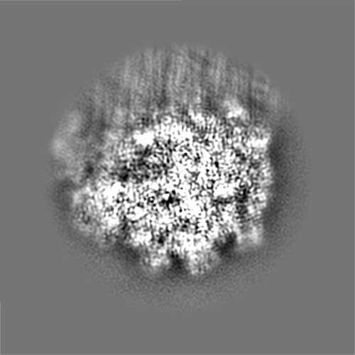
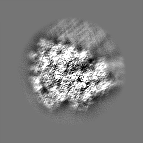
| Projections & slices | Image control
Images are generated by Spider. | ||||||||||||||||||||||||||||||||||||
| Voxel size | X=Y=Z: 0.665 Å | ||||||||||||||||||||||||||||||||||||
| Density |
| ||||||||||||||||||||||||||||||||||||
| Symmetry | Space group: 1 | ||||||||||||||||||||||||||||||||||||
| Details | EMDB XML:
|
 Z (Sec.)
 Y (Row.)
 X (Col.)


-Supplemental data
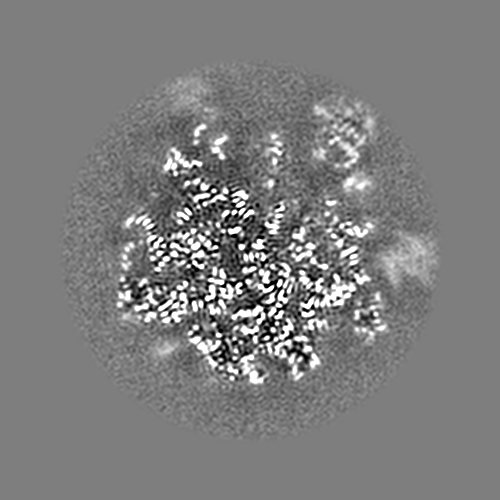
-Half map: #1
| File | emd_26657_half_map_1.map | ||||||||||||
|---|---|---|---|---|---|---|---|---|---|---|---|---|---|
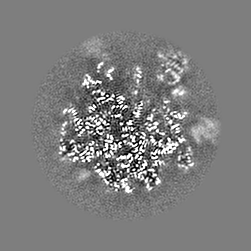
| Projections & Slices |
| ||||||||||||
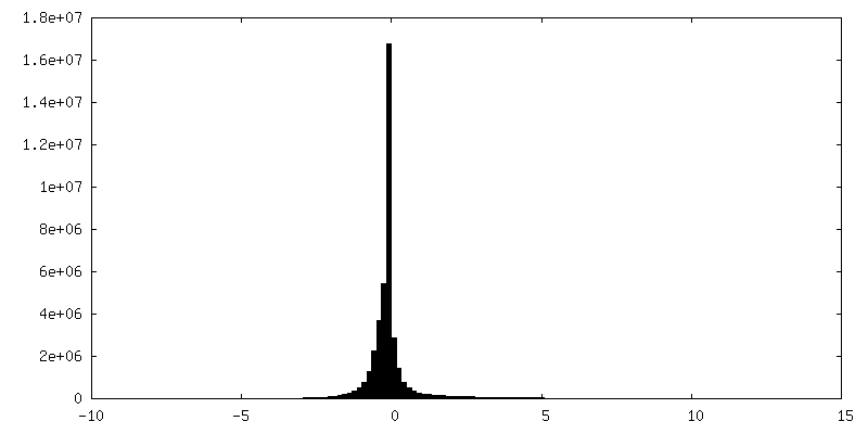
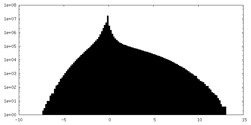
| Density Histograms |


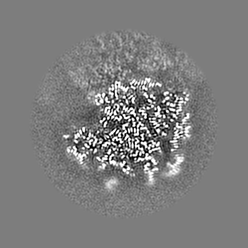

-Half map: #2
| File | emd_26657_half_map_2.map | ||||||||||||
|---|---|---|---|---|---|---|---|---|---|---|---|---|---|
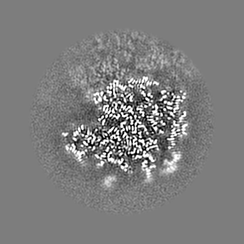
| Projections & Slices |
| ||||||||||||
| Density Histograms |


- Sample components
-Entire : 50S E. coli ribosome bound to streptogramins VM2 and VS1
| Entire | Name: 50S E. coli ribosome bound to streptogramins VM2 and VS1 |
|---|---|
| Components |
|
-Supramolecule #1: 50S E. coli ribosome bound to streptogramins VM2 and VS1
| Supramolecule | Name: 50S E. coli ribosome bound to streptogramins VM2 and VS1 type: complex / ID: 1 / Parent: 0 |
|---|---|
| Source (natural) | Organism: |
-Experimental details
-Structure determination
| Method | cryo EM |
|---|---|
 Processing Processing | single particle reconstruction |
| Aggregation state | particle |
-Sample preparation
| Buffer | pH: 7.5 |
|---|---|
| Vitrification | Cryogen name: ETHANE |
- Electron microscopy
| Microscope | FEI TITAN KRIOS |
|---|---|
| Image recording | Film or detector model: GATAN K3 (6k x 4k) / Average electron dose: 69.0 e/Å2 |
| Electron beam | Acceleration voltage: 300 kV / Electron source:  FIELD EMISSION GUN FIELD EMISSION GUN |
| Electron optics | Illumination mode: FLOOD BEAM / Imaging mode: BRIGHT FIELD / Cs: 2.7 mm / Nominal defocus max: 1.0 µm / Nominal defocus min: 0.4 µm / Nominal magnification: 130000 |
| Experimental equipment |  Model: Titan Krios / Image courtesy: FEI Company |
-Image processing
| Startup model | Type of model: NONE |
|---|---|
| Final reconstruction | Resolution.type: BY AUTHOR / Resolution: 2.29 Å / Resolution method: FSC 0.143 CUT-OFF / Number images used: 134924 |
| Initial angle assignment | Type: PROJECTION MATCHING |
| Final angle assignment | Type: PROJECTION MATCHING |
