National Institutes of Health/National Institute of General Medical Sciences (NIH/NIGMS)
GM136508
米国
引用
ジャーナル: J Struct Biol X / 年: 2022 タイトル: Hydrogens and hydrogen-bond networks in macromolecular MicroED data. 著者: Max T B Clabbers / Michael W Martynowycz / Johan Hattne / Tamir Gonen / 要旨: Microcrystal electron diffraction (MicroED) is a powerful technique utilizing electron cryo-microscopy (cryo-EM) for protein structure determination of crystalline samples too small for X-ray ...Microcrystal electron diffraction (MicroED) is a powerful technique utilizing electron cryo-microscopy (cryo-EM) for protein structure determination of crystalline samples too small for X-ray crystallography. Electrons interact with the electrostatic potential of the sample, which means that the scattered electrons carry information about the charged state of atoms and provide relatively stronger contrast for visualizing hydrogen atoms. Accurately identifying the positions of hydrogen atoms, and by extension the hydrogen bonding networks, is of importance for understanding protein structure and function, in particular for drug discovery. However, identification of individual hydrogen atom positions typically requires atomic resolution data, and has thus far remained elusive for macromolecular MicroED. Recently, we presented the structure of triclinic hen egg-white lysozyme at 0.87 Å resolution. The corresponding data were recorded under low exposure conditions using an electron-counting detector from thin crystalline lamellae. Here, using these subatomic resolution MicroED data, we identified over a third of all hydrogen atom positions based on strong difference peaks, and directly visualize hydrogen bonding interactions and the charged states of residues. Furthermore, we find that the hydrogen bond lengths are more accurately described by the inter-nuclei distances than the centers of mass of the corresponding electron clouds. We anticipate that MicroED, coupled with ongoing advances in data collection and refinement, can open further avenues for structural biology by uncovering the hydrogen atoms and hydrogen bonding interactions underlying protein structure and function.
 ムービー
ムービー コントローラー


 データを開く
 基本情報
基本情報
 マップデータ
 試料
試料 キーワード
キーワード 機能・相同性情報
 データ登録者
 米国, 2件
  引用
引用 構造の表示
構造の表示
 ダウンロードとリンク
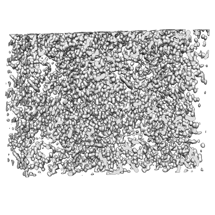 emd_26596.png
 http://ftp.pdbj.org/pub/emdb/structures/EMD-26596


 X (Sec.)
 Y (Row.)
 Z (Col.)


 試料の構成要素

 解析
解析 電子顕微鏡法
 FIELD EMISSION GUN

