National Institutes of Health/National Institute of Biomedical Imaging and Bioengineering (NIH/NIBIB)
GM122588
United States
Austrian Science Fund
P33367
Austria
Citation
Journal: Curr Biol / Year: 2022 Title: Cryo-electron tomography of the onion cell wall shows bimodally oriented cellulose fibers and reticulated homogalacturonan networks. Authors: William J Nicolas / Florian Fäßler / Przemysław Dutka / Florian K M Schur / Grant Jensen / Elliot Meyerowitz / Abstract: One hallmark of plant cells is their cell wall. They protect cells against the environment and high turgor and mediate morphogenesis through the dynamics of their mechanical and chemical properties. ...One hallmark of plant cells is their cell wall. They protect cells against the environment and high turgor and mediate morphogenesis through the dynamics of their mechanical and chemical properties. The walls are a complex polysaccharidic structure. Although their biochemical composition is well known, how the different components organize in the volume of the cell wall and interact with each other is not well understood and yet is key to the wall's mechanical properties. To investigate the ultrastructure of the plant cell wall, we imaged the walls of onion (Allium cepa) bulbs in a near-native state via cryo-focused ion beam milling (cryo-FIB milling) and cryo-electron tomography (cryo-ET). This allowed the high-resolution visualization of cellulose fibers in situ. We reveal the coexistence of dense fiber fields bathed in a reticulated matrix we termed "meshing," which is more abundant at the inner surface of the cell wall. The fibers adopted a regular bimodal angular distribution at all depths in the cell wall and bundled according to their orientation, creating layers within the cell wall. Concomitantly, employing homogalacturonan (HG)-specific enzymatic digestion, we observed changes in the meshing, suggesting that it is-at least in part-composed of HG pectins. We propose the following model for the construction of the abaxial epidermal primary cell wall: the cell deposits successive layers of cellulose fibers at -45° and +45° relative to the cell's long axis and secretes the surrounding HG-rich meshing proximal to the plasma membrane, which then migrates to more distal regions of the cell wall.
In the structure databanks used in Yorodumi, some data are registered as the other names, "COVID-19 virus" and "2019-nCoV". Here are the details of the virus and the list of structure data.
Jan 31, 2019. EMDB accession codes are about to change! (news from PDBe EMDB page)
EMDB accession codes are about to change! (news from PDBe EMDB page)
The allocation of 4 digits for EMDB accession codes will soon come to an end. Whilst these codes will remain in use, new EMDB accession codes will include an additional digit and will expand incrementally as the available range of codes is exhausted. The current 4-digit format prefixed with “EMD-” (i.e. EMD-XXXX) will advance to a 5-digit format (i.e. EMD-XXXXX), and so on. It is currently estimated that the 4-digit codes will be depleted around Spring 2019, at which point the 5-digit format will come into force.
The EM Navigator/Yorodumi systems omit the EMD- prefix.
Related info.:Q: What is EMD? / ID/Accession-code notation in Yorodumi/EM Navigator
Yorodumi is a browser for structure data from EMDB, PDB, SASBDB, etc.
This page is also the successor to EM Navigator detail page, and also detail information page/front-end page for Omokage search.
The word "yorodu" (or yorozu) is an old Japanese word meaning "ten thousand". "mi" (miru) is to see.
Related info.:EMDB / PDB / SASBDB / Comparison of 3 databanks / Yorodumi Search / Aug 31, 2016. New EM Navigator & Yorodumi / Yorodumi Papers / Jmol/JSmol / Function and homology information / Changes in new EM Navigator and Yorodumi
 Movie
Movie Controller

 Yorodumi
 Open data
 Basic information
 Map data
 Sample
 Authors
 United States,
  Austria, 3 items
  Citation
 Structure visualization
 Downloads & links
 EMDB map data format
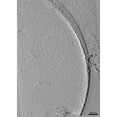 emd_26570.png
 http://ftp.pdbj.org/pub/emdb/structures/EMD-26570


 Z (Sec.)
 Y (Row.)
 X (Col.)


 Sample components
 Processing
 Electron microscopy
 FIELD EMISSION GUN
