National Institutes of Health/National Institute Of Allergy and Infectious Diseases (NIH/NIAID)
AI094386
United States
National Institutes of Health/National Institute of General Medical Sciences (NIH/NIGMS)
GM071940
United States
National Institutes of Health/Office of the Director
1S10OD018111
United States
National Institutes of Health/National Institute of General Medical Sciences (NIH/NIGMS)
1U24GM116792
United States
Citation
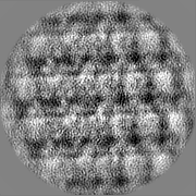
Journal: Proc Natl Acad Sci U S A / Year: 2022 Title: Locations and in situ structure of the polymerase complex inside the virion of vesicular stomatitis virus. Authors: Zhu Si / Kang Zhou / Jun Tsao / Ming Luo / Z Hong Zhou / Abstract: The polymerase complex of nonsegmented negative-strand RNA viruses primarily consists of a large (L) protein and a phosphoprotein (P). L is a multifunctional enzyme carrying out RNA-dependent RNA ...The polymerase complex of nonsegmented negative-strand RNA viruses primarily consists of a large (L) protein and a phosphoprotein (P). L is a multifunctional enzyme carrying out RNA-dependent RNA polymerization and all other steps associated with transcription and replication, while P is the nonenzymatic cofactor, regulating the function and conformation of L. The structure of a purified vesicular stomatitis virus (VSV) polymerase complex containing L and associated P segments has been determined; however, the location and manner of the attachments of L and P within each virion are unknown, limiting our mechanistic understanding of VSV RNA replication and transcription and hindering engineering efforts of this widely used anticancer and vaccine vector. Here, we have used cryo-electron tomography to visualize the VSV virion, revealing the attachment of the ring-shaped L molecules to VSV nucleocapsid proteins (N) throughout the cavity of the bullet-shaped nucleocapsid. Subtomogram averaging and three-dimensional classification of regions containing N and the matrix protein (M) have yielded the in situ structure of the polymerase complex. On average, ∼55 polymerase complexes are packaged in each virion. The capping domain of L interacts with two neighboring N molecules through flexible attachments. P, which exists as a dimer, bridges separate N molecules and the connector and C-terminal domains of L. Our data provide the structural basis for recruitment of L to N by P in virus assembly and for flexible attachments between L and N, which allow a quick response of L in primary transcription upon cell entry.
In the structure databanks used in Yorodumi, some data are registered as the other names, "COVID-19 virus" and "2019-nCoV". Here are the details of the virus and the list of structure data.
Jan 31, 2019. EMDB accession codes are about to change! (news from PDBe EMDB page)
EMDB accession codes are about to change! (news from PDBe EMDB page)
The allocation of 4 digits for EMDB accession codes will soon come to an end. Whilst these codes will remain in use, new EMDB accession codes will include an additional digit and will expand incrementally as the available range of codes is exhausted. The current 4-digit format prefixed with “EMD-” (i.e. EMD-XXXX) will advance to a 5-digit format (i.e. EMD-XXXXX), and so on. It is currently estimated that the 4-digit codes will be depleted around Spring 2019, at which point the 5-digit format will come into force.
The EM Navigator/Yorodumi systems omit the EMD- prefix.
Related info.:Q: What is EMD? / ID/Accession-code notation in Yorodumi/EM Navigator
Yorodumi is a browser for structure data from EMDB, PDB, SASBDB, etc.
This page is also the successor to EM Navigator detail page, and also detail information page/front-end page for Omokage search.
The word "yorodu" (or yorozu) is an old Japanese word meaning "ten thousand". "mi" (miru) is to see.
Related info.:EMDB / PDB / SASBDB / Comparison of 3 databanks / Yorodumi Search / Aug 31, 2016. New EM Navigator & Yorodumi / Yorodumi Papers / Jmol/JSmol / Function and homology information / Changes in new EM Navigator and Yorodumi
 Movie
Movie Controller


 Open data
 Basic information
 Map data
 Sample
 Vesicular stomatitis Indiana virus strain San Juan
 Authors
 United States, 4 items
  Citation
 Structure visualization
 Downloads & links
 EMDB map data format
 emd_26465.png
 http://ftp.pdbj.org/pub/emdb/structures/EMD-26465
 Z (Sec.)
 Y (Row.)
 X (Col.)


 Sample components
 Homo sapiens (human) / Recombinant cell: HeLa
 Processing
 Electron microscopy
 FIELD EMISSION GUN