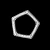
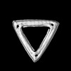
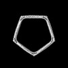
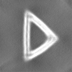
Journal: Sci Adv / Year: 2022 Title: Planar 2D wireframe DNA origami. Authors: Xiao Wang / Shanshan Li / Hyungmin Jun / Torsten John / Kaiming Zhang / Hannah Fowler / Jonathan P K Doye / Wah Chiu / Mark Bathe / Abstract: Two-dimensional (2D) DNA origami is widely used for applications ranging from excitonics to single-molecule biophysics. Conventional, single-layer 2D DNA origami exhibits flexibility and curvature in ...Two-dimensional (2D) DNA origami is widely used for applications ranging from excitonics to single-molecule biophysics. Conventional, single-layer 2D DNA origami exhibits flexibility and curvature in solution; however, that may limit its suitability as a 2D structural template. In contrast, 2D wireframe DNA origami rendered with six-helix bundle edges offers local control over duplex orientations with enhanced in-plane rigidity. Here, we investigate the 3D structure of these assemblies using cryo-electron microscopy (cryo-EM). 3D reconstructions reveal a high degree of planarity and homogeneity in solution for polygonal objects with and without internal mesh, enabling 10-Å resolution for a triangle. Coarse-grained simulations were in agreement with cryo-EM data, offering molecular structural insight into this class of 2D DNA origami. Our results suggest that these assemblies may be valuable for 2D material applications and geometries that require high structural fidelity together with local control over duplex orientations, rather than parallel duplex assembly.
In the structure databanks used in Yorodumi, some data are registered as the other names, "COVID-19 virus" and "2019-nCoV". Here are the details of the virus and the list of structure data.
Jan 31, 2019. EMDB accession codes are about to change! (news from PDBe EMDB page)
EMDB accession codes are about to change! (news from PDBe EMDB page)
The allocation of 4 digits for EMDB accession codes will soon come to an end. Whilst these codes will remain in use, new EMDB accession codes will include an additional digit and will expand incrementally as the available range of codes is exhausted. The current 4-digit format prefixed with “EMD-” (i.e. EMD-XXXX) will advance to a 5-digit format (i.e. EMD-XXXXX), and so on. It is currently estimated that the 4-digit codes will be depleted around Spring 2019, at which point the 5-digit format will come into force.
The EM Navigator/Yorodumi systems omit the EMD- prefix.
Related info.:Q: What is EMD? / ID/Accession-code notation in Yorodumi/EM Navigator
Yorodumi is a browser for structure data from EMDB, PDB, SASBDB, etc.
This page is also the successor to EM Navigator detail page, and also detail information page/front-end page for Omokage search.
The word "yorodu" (or yorozu) is an old Japanese word meaning "ten thousand". "mi" (miru) is to see.
Related info.:EMDB / PDB / SASBDB / Comparison of 3 databanks / Yorodumi Search / Aug 31, 2016. New EM Navigator & Yorodumi / Yorodumi Papers / Jmol/JSmol / Function and homology information / Changes in new EM Navigator and Yorodumi
 Movie
Movie Controller


 Open data
 Basic information
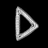
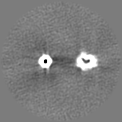
 Map data
 Sample
 Escherichia virus M13
 Authors
 United States, 4 items
  Citation


 Structure visualization
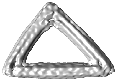
 Downloads & links
 EMDB map data format
 emd_26328.png
 http://ftp.pdbj.org/pub/emdb/structures/EMD-26328
 Z (Sec.)
 Y (Row.)
 X (Col.)


 Sample components
 Processing
 Electron microscopy
 FIELD EMISSION GUN
