 Movie
Movie Controller


+ Open data

- Basic information
| Entry |  | |||||||||
|---|---|---|---|---|---|---|---|---|---|---|
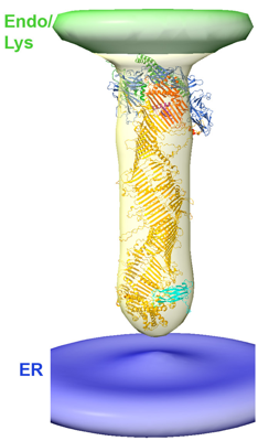
| Title | In situ architecture of VPS13C | |||||||||
 Map data Map data | ||||||||||
 Sample Sample |
| |||||||||
 Keywords Keywords | Lipid transfer / LIPID TRANSPORT | |||||||||
| Biological species |  Homo sapiens (human) Homo sapiens (human) | |||||||||
| Method | subtomogram averaging / cryo EM / Resolution: 47.0 Å | |||||||||
 Authors Authors | Cai S / De Camilli P | |||||||||
| Funding support |  United States, 2 items United States, 2 items
| |||||||||
 Citation Citation | Journal: Proc Natl Acad Sci U S A / Year: 2022 Title: In situ architecture of the lipid transport protein VPS13C at ER-lysosome membrane contacts. Authors: Shujun Cai / Yumei Wu / Andrés Guillén-Samander / William Hancock-Cerutti / Jun Liu / Pietro De Camilli / Abstract: VPS13 is a eukaryotic lipid transport protein localized at membrane contact sites. Previous studies suggested that it may transfer lipids between adjacent bilayers by a bridge-like mechanism. Direct ...VPS13 is a eukaryotic lipid transport protein localized at membrane contact sites. Previous studies suggested that it may transfer lipids between adjacent bilayers by a bridge-like mechanism. Direct evidence for this hypothesis from a full-length structure and from electron microscopy (EM) studies in situ is still missing, however. Here, we have capitalized on AlphaFold predictions to complement the structural information already available about VPS13 and to generate a full-length model of human VPS13C, the Parkinson's disease-linked VPS13 paralog localized at contacts between the endoplasmic reticulum (ER) and endo/lysosomes. Such a model predicts an ∼30-nm rod with a hydrophobic groove that extends throughout its length. We further investigated whether such a structure can be observed in situ at ER-endo/lysosome contacts. To this aim, we combined genetic approaches with cryo-focused ion beam (cryo-FIB) milling and cryo-electron tomography (cryo-ET) to examine HeLa cells overexpressing this protein (either full length or with an internal truncation) along with VAP, its anchoring binding partner at the ER. Using these methods, we identified rod-like densities that span the space separating the two adjacent membranes and that match the predicted structures of either full-length VPS13C or its shorter truncated mutant, thus providing in situ evidence for a bridge model of VPS13 in lipid transport. | |||||||||
| History |
|
- Structure visualization
| Supplemental images |
|---|
- Downloads & links
-EMDB archive
| Map data | emd_26247.map.gz | 859.6 KB |  EMDB map data format EMDB map data format | |
|---|---|---|---|---|
| Header (meta data) | emd-26247-v30.xmlemd-26247.xml | 12.4 KB 12.4 KB | Display Display | EMDB header |
| Images |  emd_26247.png emd_26247.png | 78.3 KB | ||
| Filedesc metadata | emd-26247.cif.gz | 3.8 KB | ||
| Others | emd_26247_additional_1.map.gzemd_26247_additional_2.map.gz | 947.3 KB 687.3 KB | ||
| Archive directory |  http://ftp.pdbj.org/pub/emdb/structures/EMD-26247ftp://ftp.pdbj.org/pub/emdb/structures/EMD-26247 http://ftp.pdbj.org/pub/emdb/structures/EMD-26247ftp://ftp.pdbj.org/pub/emdb/structures/EMD-26247 | HTTPS FTP |
-Related structure data
| Related structure data | |
|---|---|
| EM raw data | EMPIAR-10962 (Title: In situ architecture of the lipid transport protein VPS13C at ER-lysosomes membrane contacts Data size: 29.5 Data #1: Tilt series and tomograms used in a cryo-ET study of in situ VPS13C [tilt series]) |

-Links
| EMDB pages | EMDB (EBI/PDBe) / EMDataResource |
|---|
-Map
| File | Download / File: emd_26247.map.gz / Format: CCP4 / Size: 1 MB / Type: IMAGE STORED AS FLOATING POINT NUMBER (4 BYTES) | ||||||||||||||||||||||||||||||||||||
|---|---|---|---|---|---|---|---|---|---|---|---|---|---|---|---|---|---|---|---|---|---|---|---|---|---|---|---|---|---|---|---|---|---|---|---|---|---|
| Projections & slices | Image control
Images are generated by Spider. | ||||||||||||||||||||||||||||||||||||
| Voxel size | X=Y=Z: 13.536 Å | ||||||||||||||||||||||||||||||||||||
| Density |
| ||||||||||||||||||||||||||||||||||||
| Symmetry | Space group: 1 | ||||||||||||||||||||||||||||||||||||
| Details | EMDB XML:
|
 Z (Sec.)
 Y (Row.)
 X (Col.)


-Supplemental data
-Additional map: #1
| File | emd_26247_additional_1.map | ||||||||||||
|---|---|---|---|---|---|---|---|---|---|---|---|---|---|
| Projections & Slices |
| ||||||||||||
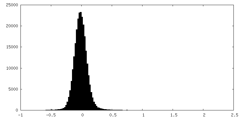
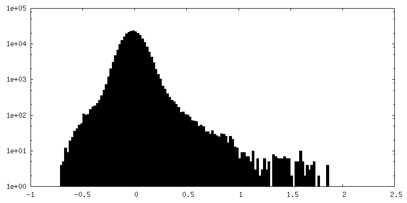
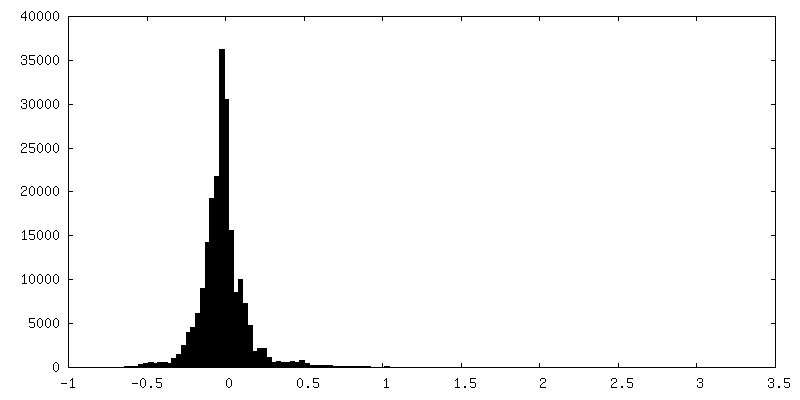
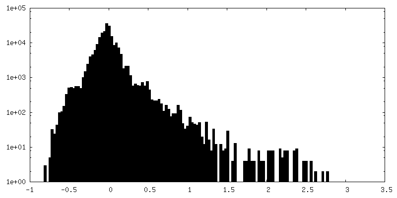
| Density Histograms |


-Additional map: #2
| File | emd_26247_additional_2.map | ||||||||||||
|---|---|---|---|---|---|---|---|---|---|---|---|---|---|
| Projections & Slices |
| ||||||||||||
| Density Histograms |


- Sample components
-Entire : full-length human VPS13C bridging the two adjacent membranes
| Entire | Name: full-length human VPS13C bridging the two adjacent membranes |
|---|---|
| Components |
|
-Supramolecule #1: full-length human VPS13C bridging the two adjacent membranes
| Supramolecule | Name: full-length human VPS13C bridging the two adjacent membranes type: cell / ID: 1 / Parent: 0 Details: Subtomogram-average density maps showing a full-length VPS13C rod bridging the two adjacent membranes. C100 symmetry was applied to enhance signal to noise ratio. |
|---|---|
| Source (natural) | Organism: Homo sapiens (human) |
-Experimental details
-Structure determination
| Method | cryo EM |
|---|---|
 Processing Processing | subtomogram averaging |
| Aggregation state | cell |
-Sample preparation
| Buffer | pH: 7.4 |
|---|---|
| Vitrification | Cryogen name: ETHANE-PROPANE |
- Electron microscopy
| Microscope | FEI TITAN KRIOS |
|---|---|
| Image recording | Film or detector model: GATAN K3 (6k x 4k) / Average electron dose: 2.0 e/Å2 |
| Electron beam | Acceleration voltage: 300 kV / Electron source:  FIELD EMISSION GUN FIELD EMISSION GUN |
| Electron optics | Illumination mode: FLOOD BEAM / Imaging mode: BRIGHT FIELD / Nominal defocus max: -0.006 µm / Nominal defocus min: -0.001 µm |
| Experimental equipment |  Model: Titan Krios / Image courtesy: FEI Company |
-Image processing
| Final reconstruction | Algorithm: BACK PROJECTION / Resolution.type: BY AUTHOR / Resolution: 47.0 Å / Resolution method: FSC 0.143 CUT-OFF / Software - Name: PROTOMO / Number subtomograms used: 570 |
|---|---|
| Extraction | Number tomograms: 20 / Number images used: 570 |
| Final angle assignment | Type: NOT APPLICABLE |
