Movie
Movie Controller


+ Open data

- Basic information
| Entry |  | |||||||||
|---|---|---|---|---|---|---|---|---|---|---|
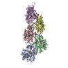
| Title | F-actin from neuronal growth cone filopodium | |||||||||
 Map data Map data | Subtomogram average of filamentous actin from a neuronal growth cone filopodium. | |||||||||
 Sample Sample |
| |||||||||
 Keywords Keywords | Cytoskeleton / Polymer / Filament / Actin / STRUCTURAL PROTEIN | |||||||||
| Biological species |  | |||||||||
| Method | subtomogram averaging / cryo EM / Resolution: 20.2 Å | |||||||||
 Authors Authors | Hylton RK / Swulius MT | |||||||||
| Funding support | 1 items
| |||||||||
 Citation Citation | Journal: Nat Commun / Year: 2022 Title: Cofilactin filaments regulate filopodial structure and dynamics in neuronal growth cones. Authors: Ryan K Hylton / Jessica E Heebner / Michael A Grillo / Matthew T Swulius /  Abstract: Cofilin is best known for its ability to sever actin filaments and facilitate cytoskeletal recycling inside of cells, but at higher concentrations in vitro, cofilin stabilizes a more flexible, hyper- ...Cofilin is best known for its ability to sever actin filaments and facilitate cytoskeletal recycling inside of cells, but at higher concentrations in vitro, cofilin stabilizes a more flexible, hyper-twisted state of actin known as "cofilactin". While this filament state is well studied, a structural role for cofilactin in dynamic cellular processes has not been observed. With a combination of cryo-electron tomography and fluorescence imaging in neuronal growth cones, we observe that filopodial actin filaments switch between a fascin-linked and a cofilin-decorated state, and that cofilactin is associated with a variety of dynamic events within filopodia. The switch to cofilactin filaments occurs in a graded fashion and correlates with a decline in fascin cross-linking within the filopodia, which is associated with curvature in the bundle. Our tomographic data reveal that the hyper-twisting of actin from cofilin binding leads to a rearrangement of filament packing, which largely excludes fascin from the base of filopodia. Our results provide mechanistic insight into the fundamentals of cytoskeletal remodeling inside of confined cellular spaces, and how the interplay between fascin and cofilin regulates the dynamics of searching filopodia. | |||||||||
| History |
|
- Structure visualization
| Supplemental images |
|---|

- Downloads & links
-EMDB archive
| Map data | emd_26214.map.gz | 1.6 MB |  EMDB map data format EMDB map data format | |
|---|---|---|---|---|
| Header (meta data) | emd-26214-v30.xmlemd-26214.xml | 11.6 KB 11.6 KB | Display Display | EMDB header |
| Images |  emd_26214.png emd_26214.png | 43 KB | ||
| Filedesc metadata | emd-26214.cif.gz | 4.6 KB | ||
| Archive directory |  http://ftp.pdbj.org/pub/emdb/structures/EMD-26214ftp://ftp.pdbj.org/pub/emdb/structures/EMD-26214 http://ftp.pdbj.org/pub/emdb/structures/EMD-26214ftp://ftp.pdbj.org/pub/emdb/structures/EMD-26214 | HTTPS FTP |
-Related structure data

-Links
| EMDB pages | EMDB (EBI/PDBe) / EMDataResource |
|---|
-Map
| File | Download / File: emd_26214.map.gz / Format: CCP4 / Size: 1.7 MB / Type: IMAGE STORED AS FLOATING POINT NUMBER (4 BYTES) | ||||||||||||||||||||||||||||||||||||
|---|---|---|---|---|---|---|---|---|---|---|---|---|---|---|---|---|---|---|---|---|---|---|---|---|---|---|---|---|---|---|---|---|---|---|---|---|---|
| Annotation | Subtomogram average of filamentous actin from a neuronal growth cone filopodium. | ||||||||||||||||||||||||||||||||||||
| Projections & slices | Image control
Images are generated by Spider. generated in cubic-lattice coordinate | ||||||||||||||||||||||||||||||||||||
| Voxel size | X=Y=Z: 4.306 Å | ||||||||||||||||||||||||||||||||||||
| Density |
| ||||||||||||||||||||||||||||||||||||
| Symmetry | Space group: 1 | ||||||||||||||||||||||||||||||||||||
| Details | EMDB XML:
|
 Z (Sec.)
 Y (Row.)
 X (Col.)


-Supplemental data
- Sample components
-Entire : Filamentous actin from a neuronal growth cone filopodium
| Entire | Name: Filamentous actin from a neuronal growth cone filopodium |
|---|---|
| Components |
|
-Supramolecule #1: Filamentous actin from a neuronal growth cone filopodium
| Supramolecule | Name: Filamentous actin from a neuronal growth cone filopodium type: organelle_or_cellular_component / ID: 1 / Parent: 0 Details: Cryo-electron tomography was performed on filopodia of neurons growing on EM grids. The map is a result from subtomogram averaging of ~83 nm-long filament sections (1603 particles total) ...Details: Cryo-electron tomography was performed on filopodia of neurons growing on EM grids. The map is a result from subtomogram averaging of ~83 nm-long filament sections (1603 particles total) from an f-actin bundle in a single filopodium. |
|---|---|
| Source (natural) | Organism: |
-Experimental details
-Structure determination
| Method | cryo EM |
|---|---|
 Processing Processing | subtomogram averaging |
| Aggregation state | cell |
-Sample preparation
| Buffer | pH: 7.4 Details: No buffer was used. The cells were grown on EM grids in Neurobasal media with B-27 supplement (2%) and Penicillin/Streptomycin (1%) or NbActiv4 (with 1% Penicillin/Streptomycin) neuronal cell culture media. |
|---|---|
| Grid | Model: Quantifoil R2/2 / Material: GOLD / Mesh: 200 / Support film - Material: CARBON / Support film - topology: HOLEY / Pretreatment - Type: GLOW DISCHARGE / Pretreatment - Time: 60 sec. |
| Vitrification | Cryogen name: ETHANE / Chamber humidity: 70 % / Chamber temperature: 295.15 K / Instrument: FEI VITROBOT MARK IV Details: 3 uL of 10 nm gold fiducials were added on top of the cells prior to blotting or freezing. Grids were then blotted by hand from behind (the non-cell surface) for 2 seconds before plunge-freezing.. |
- Electron microscopy
| Microscope | FEI TITAN KRIOS |
|---|---|
| Specialist optics | Energy filter - Name: GIF Bioquantum / Energy filter - Slit width: 20 eV |
| Image recording | Film or detector model: GATAN K2 SUMMIT (4k x 4k) / Detector mode: COUNTING / Average exposure time: 0.9 sec. / Average electron dose: 2.5 e/Å2 |
| Electron beam | Acceleration voltage: 300 kV / Electron source:  FIELD EMISSION GUN FIELD EMISSION GUN |
| Electron optics | Illumination mode: FLOOD BEAM / Imaging mode: BRIGHT FIELD / Nominal defocus max: 8.0 µm / Nominal defocus min: 6.0 µm / Nominal magnification: 26000 |
| Sample stage | Specimen holder model: FEI TITAN KRIOS AUTOGRID HOLDER / Cooling holder cryogen: NITROGEN |
| Experimental equipment |  Model: Titan Krios / Image courtesy: FEI Company |
-Image processing
| Final reconstruction | Applied symmetry - Helical parameters - Δz: 27.6 Å Applied symmetry - Helical parameters - Δ&Phi: -166.6 ° Applied symmetry - Helical parameters - Axial symmetry: C1 (asymmetric) Resolution.type: BY AUTHOR / Resolution: 20.2 Å / Resolution method: FSC 0.143 CUT-OFF / Software - Name: Dynamo / Number subtomograms used: 1603 |
|---|---|
| Extraction | Number tomograms: 1 / Number images used: 1603 / Reference model: Non-refined average of all particles. / Method: Filaments picked by hand. / Software - Name: Dynamo |
| Final angle assignment | Type: NOT APPLICABLE |
-Atomic model buiding 1
| Initial model | PDB ID: Chain - Source name: PDB / Chain - Initial model type: experimental model |
|---|---|
| Refinement | Protocol: RIGID BODY FIT |