[English] 日本語
 Yorodumi
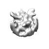
- EMDB-26189: Third class of AAV5 bound with PKD1-2 revealed by classification ... -
+ Open data
- Basic information
| Entry |  | |||||||||
|---|---|---|---|---|---|---|---|---|---|---|
| Title | Third class of AAV5 bound with PKD1-2 revealed by classification of aligned subtomograms | |||||||||
 Map data Map data | Third class of AAV5 bound with PKD1-2 revealed by classification of aligned subtomgrams | |||||||||
 Sample Sample |
| |||||||||
| Biological species |  adeno-associated virus 5 / adeno-associated virus 5 /  Homo sapiens (human) Homo sapiens (human) | |||||||||
| Method | subtomogram averaging / cryo EM / Resolution: 25.0 Å | |||||||||
 Authors Authors | Hu GQ / Silveria MA / Chapman MS / Stagg SM | |||||||||
| Funding support |  United States, 2 items United States, 2 items
| |||||||||
 Citation Citation |  Journal: J Virol / Year: 2022 Journal: J Virol / Year: 2022Title: Adeno-Associated Virus Receptor-Binding: Flexible Domains and Alternative Conformations through Cryo-Electron Tomography of Adeno-Associated Virus 2 (AAV2) and AAV5 Complexes. Authors: Guiqing Hu / Mark A Silveria / Grant M Zane / Michael S Chapman / Scott M Stagg /  Abstract: Recombinant forms of adeno-associated virus (rAAV) are vectors of choice in the development of treatments for a number of genetic dispositions. Greater understanding of AAV's molecular virology is ...Recombinant forms of adeno-associated virus (rAAV) are vectors of choice in the development of treatments for a number of genetic dispositions. Greater understanding of AAV's molecular virology is needed to underpin needed improvements in efficiency and specificity. Recent advances have included identification of a near-universal entry receptor, AAVR, and structures detected by cryo-electron microscopy (EM) single particle analysis (SPA) that revealed, at high resolution, only the domains of AAVR most tightly bound to AAV. Here, cryogenic electron tomography (cryo-ET) is applied to reveal the neighboring domains of the flexible receptor. For AAV5, where the PKD1 domain is bound strongly, PKD2 is seen in three configurations extending away from the virus. AAV2 binds tightly to the PKD2 domain at a distinct site, and cryo-ET now reveals four configurations of PKD1, all different from that seen in AAV5. The AAV2 receptor complex also shows unmodeled features on the inner surface that appear to be an equilibrium alternate configuration. Other AAV structures start near the 5-fold axis, but now β-strand A is the minor conformer and, for the major conformer, partially ordered N termini near the 2-fold axis join the canonical capsid jellyroll fold at the βA-βB turn. The addition of cryo-ET is revealing unappreciated complexity that is likely relevant to viral entry and to the development of improved gene therapy vectors. With 150 clinical trials for 30 diseases under way, AAV is a leading gene therapy vector. Immunotoxicity at high doses used to overcome inefficient transduction has occasionally proven fatal and highlighted gaps in fundamental virology. AAV enters cells, interacting through distinct sites with different domains of the AAVR receptor, according to AAV clade. Single domains are resolved in structures by cryogenic electron microscopy. Here, the adjoining domains are revealed by cryo-electron tomography of AAV2 and AAV5 complexes. They are in flexible configurations interacting minimally with AAV, despite measurable dependence of AAV2 transduction on both domains. | |||||||||
| History |
|
- Structure visualization
| Supplemental images |
|---|
- Downloads & links
-EMDB archive
| Map data |  emd_26189.map.gz emd_26189.map.gz | 32.7 KB |  EMDB map data format EMDB map data format | |
|---|---|---|---|---|
| Header (meta data) |  emd-26189-v30.xml emd-26189-v30.xml emd-26189.xml emd-26189.xml | 13.3 KB 13.3 KB | Display Display |  EMDB header EMDB header |
| Images |  emd_26189.png emd_26189.png | 44.6 KB | ||
| Archive directory |  http://ftp.pdbj.org/pub/emdb/structures/EMD-26189 http://ftp.pdbj.org/pub/emdb/structures/EMD-26189 ftp://ftp.pdbj.org/pub/emdb/structures/EMD-26189 ftp://ftp.pdbj.org/pub/emdb/structures/EMD-26189 | HTTPS FTP |
-Validation report
| Summary document |  emd_26189_validation.pdf.gz emd_26189_validation.pdf.gz | 313 KB | Display |  EMDB validaton report EMDB validaton report |
|---|---|---|---|---|
| Full document |  emd_26189_full_validation.pdf.gz emd_26189_full_validation.pdf.gz | 312.6 KB | Display | |
| Data in XML |  emd_26189_validation.xml.gz emd_26189_validation.xml.gz | 4.6 KB | Display | |
| Data in CIF |  emd_26189_validation.cif.gz emd_26189_validation.cif.gz | 5.2 KB | Display | |
| Arichive directory |  https://ftp.pdbj.org/pub/emdb/validation_reports/EMD-26189 https://ftp.pdbj.org/pub/emdb/validation_reports/EMD-26189 ftp://ftp.pdbj.org/pub/emdb/validation_reports/EMD-26189 ftp://ftp.pdbj.org/pub/emdb/validation_reports/EMD-26189 | HTTPS FTP |
-Related structure data
| Related structure data | C: citing same article ( |
|---|
- Links
Links
| EMDB pages |  EMDB (EBI/PDBe) / EMDB (EBI/PDBe) /  EMDataResource EMDataResource |
|---|
- Map
Map
| File |  Download / File: emd_26189.map.gz / Format: CCP4 / Size: 356.4 KB / Type: IMAGE STORED AS FLOATING POINT NUMBER (4 BYTES) Download / File: emd_26189.map.gz / Format: CCP4 / Size: 356.4 KB / Type: IMAGE STORED AS FLOATING POINT NUMBER (4 BYTES) | ||||||||||||||||||||||||||||||||||||
|---|---|---|---|---|---|---|---|---|---|---|---|---|---|---|---|---|---|---|---|---|---|---|---|---|---|---|---|---|---|---|---|---|---|---|---|---|---|
| Annotation | Third class of AAV5 bound with PKD1-2 revealed by classification of aligned subtomgrams | ||||||||||||||||||||||||||||||||||||
| Projections & slices | Image control
Images are generated by Spider. | ||||||||||||||||||||||||||||||||||||
| Voxel size | X=Y=Z: 5.48 Å | ||||||||||||||||||||||||||||||||||||
| Density |
| ||||||||||||||||||||||||||||||||||||
| Symmetry | Space group: 1 | ||||||||||||||||||||||||||||||||||||
| Details | EMDB XML:
|
-Supplemental data
- Sample components
-Entire : Binary complex of AAV-5 with a two domain fragment of its cellula...
| Entire | Name: Binary complex of AAV-5 with a two domain fragment of its cellular receptor, AAVR |
|---|---|
| Components |
|
-Supramolecule #1: Binary complex of AAV-5 with a two domain fragment of its cellula...
| Supramolecule | Name: Binary complex of AAV-5 with a two domain fragment of its cellular receptor, AAVR type: complex / Chimera: Yes / ID: 1 / Parent: 0 |
|---|---|
| Molecular weight | Experimental: 82 KDa |
-Supramolecule #2: AAV-5
| Supramolecule | Name: AAV-5 / type: complex / Chimera: Yes / ID: 2 / Parent: 1 |
|---|---|
| Source (natural) | Organism:  adeno-associated virus 5 adeno-associated virus 5 |
| Recombinant expression | Organism:  |
| Molecular weight | Theoretical: 60 KDa |
-Supramolecule #3: AAVR
| Supramolecule | Name: AAVR / type: complex / Chimera: Yes / ID: 3 / Parent: 1 |
|---|---|
| Source (natural) | Organism:  Homo sapiens (human) Homo sapiens (human) |
| Recombinant expression | Organism:  |
| Molecular weight | Theoretical: 22 KDa |
-Experimental details
-Structure determination
| Method | cryo EM |
|---|---|
 Processing Processing | subtomogram averaging |
| Aggregation state | particle |
- Sample preparation
| Buffer | pH: 7.4 Component:
| |||||||||
|---|---|---|---|---|---|---|---|---|---|---|
| Grid | Model: PELCO Ultrathin Carbon with Lacey Carbon / Support film - Material: CARBON / Support film - topology: LACEY / Support film - Film thickness: 3.0 nm | |||||||||
| Vitrification | Cryogen name: ETHANE / Chamber humidity: 100 % / Chamber temperature: 298 K / Instrument: FEI VITROBOT MARK IV Details: 4ul of AAV-2 at concentration 0.2 mg/ml was was applied on grid and incubated for 1.5 minutes. The grid was gently blotted on the side with filter paper, and another 4ul of PKD1-2 at ...Details: 4ul of AAV-2 at concentration 0.2 mg/ml was was applied on grid and incubated for 1.5 minutes. The grid was gently blotted on the side with filter paper, and another 4ul of PKD1-2 at concentration 1.5 mg/ml was applied and allowed to incubate for another 1.5 minutes followed by plunge-freeze using a Vitrobot Mark IV.. | |||||||||
| Details | 2 ul of 6.5 uM AAV5 VLP was added to the grid and given 2 minutes to adhere. Sample was then wicked and 2 ul of 33 uM PKD1-2 was added. Grids were then plunge frozen using an FEI Vitrobot Mark IV with a blot force of 4, time of 2 sec, temperature of 25 C, and at 100 percent humidity. |
- Electron microscopy
| Microscope | FEI TITAN KRIOS |
|---|---|
| Image recording | Film or detector model: GATAN K3 (6k x 4k) / Digitization - Dimensions - Width: 5760 pixel / Digitization - Dimensions - Height: 4092 pixel / Number grids imaged: 1 / Average electron dose: 100.0 e/Å2 |
| Electron beam | Acceleration voltage: 300 kV / Electron source:  FIELD EMISSION GUN FIELD EMISSION GUN |
| Electron optics | C2 aperture diameter: 100.0 µm / Calibrated defocus max: 5.0 µm / Calibrated defocus min: 5.0 µm / Calibrated magnification: 33000 / Illumination mode: FLOOD BEAM / Imaging mode: BRIGHT FIELD / Cs: 2.7 mm / Nominal defocus max: 5.0 µm / Nominal defocus min: 5.0 µm / Nominal magnification: 33000 |
| Sample stage | Specimen holder model: FEI TITAN KRIOS AUTOGRID HOLDER / Cooling holder cryogen: NITROGEN |
| Experimental equipment |  Model: Titan Krios / Image courtesy: FEI Company |
- Image processing
| Final reconstruction | Number classes used: 3 / Applied symmetry - Point group: C3 (3 fold cyclic) / Resolution.type: BY AUTHOR / Resolution: 25.0 Å / Resolution method: FSC 0.143 CUT-OFF / Software - Name: EMAN (ver. 2) / Number subtomograms used: 5100 |
|---|---|
| Extraction | Number tomograms: 1 / Number images used: 5100 / Software - Name: EMAN (ver. 2) |
| CTF correction | Software - Name: EMAN (ver. 2) |
| Final angle assignment | Type: OTHER |
 Movie
Movie Controller


 Z (Sec.)
 Y (Row.)
 X (Col.)


