[English] 日本語
 Yorodumi
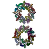
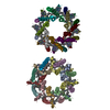
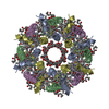
- EMDB-26134: LH2-LH3 antenna in anti parallel configuration embedded in a nanodisc -
+ Open data
- Basic information
| Entry |  | ||||||||||||||||||
|---|---|---|---|---|---|---|---|---|---|---|---|---|---|---|---|---|---|---|---|
| Title | LH2-LH3 antenna in anti parallel configuration embedded in a nanodisc | ||||||||||||||||||
 Map data Map data | LH2-LH3 antenna in anti parallel orientation with nanodisc | ||||||||||||||||||
 Sample Sample |
| ||||||||||||||||||
 Keywords Keywords | antenna / membrane protein / nanodisc / bacteriochlorophyll / PHOTOSYNTHESIS | ||||||||||||||||||
| Function / homology |  Function and homology information Function and homology informationorganelle inner membrane / plasma membrane light-harvesting complex / bacteriochlorophyll binding / : / photosynthesis, light reaction / metal ion binding / plasma membrane Similarity search - Function | ||||||||||||||||||
| Biological species |  Magnetospirillum molischianum (magnetotactic) Magnetospirillum molischianum (magnetotactic) | ||||||||||||||||||
| Method | single particle reconstruction / cryo EM / Resolution: 8.2 Å | ||||||||||||||||||
 Authors Authors | Toporik H / Harris D | ||||||||||||||||||
| Funding support |  United States, 5 items United States, 5 items
| ||||||||||||||||||
 Citation Citation |  Journal: Proc Natl Acad Sci U S A / Year: 2023 Journal: Proc Natl Acad Sci U S A / Year: 2023Title: Elucidating interprotein energy transfer dynamics within the antenna network from purple bacteria. Authors: Dihao Wang / Olivia C Fiebig / Dvir Harris / Hila Toporik / Yi Ji / Chern Chuang / Muath Nairat / Ashley L Tong / John I Ogren / Stephanie M Hart / Jianshu Cao / James N Sturgis / Yuval ...Authors: Dihao Wang / Olivia C Fiebig / Dvir Harris / Hila Toporik / Yi Ji / Chern Chuang / Muath Nairat / Ashley L Tong / John I Ogren / Stephanie M Hart / Jianshu Cao / James N Sturgis / Yuval Mazor / Gabriela S Schlau-Cohen /   Abstract: In photosynthesis, absorbed light energy transfers through a network of antenna proteins with near-unity quantum efficiency to reach the reaction center, which initiates the downstream biochemical ...In photosynthesis, absorbed light energy transfers through a network of antenna proteins with near-unity quantum efficiency to reach the reaction center, which initiates the downstream biochemical reactions. While the energy transfer dynamics within individual antenna proteins have been extensively studied over the past decades, the dynamics between the proteins are poorly understood due to the heterogeneous organization of the network. Previously reported timescales averaged over such heterogeneity, obscuring individual interprotein energy transfer steps. Here, we isolated and interrogated interprotein energy transfer by embedding two variants of the primary antenna protein from purple bacteria, light-harvesting complex 2 (LH2), together into a near-native membrane disc, known as a nanodisc. We integrated ultrafast transient absorption spectroscopy, quantum dynamics simulations, and cryogenic electron microscopy to determine interprotein energy transfer timescales. By varying the diameter of the nanodiscs, we replicated a range of distances between the proteins. The closest distance possible between neighboring LH2, which is the most common in native membranes, is 25 Å and resulted in a timescale of 5.7 ps. Larger distances of 28 to 31 Å resulted in timescales of 10 to 14 ps. Corresponding simulations showed that the fast energy transfer steps between closely spaced LH2 increase transport distances by ∼15%. Overall, our results introduce a framework for well-controlled studies of interprotein energy transfer dynamics and suggest that protein pairs serve as the primary pathway for the efficient transport of solar energy. | ||||||||||||||||||
| History |
|
- Structure visualization
| Supplemental images |
|---|
- Downloads & links
-EMDB archive
| Map data |  emd_26134.map.gz emd_26134.map.gz | 4.6 MB |  EMDB map data format EMDB map data format | |
|---|---|---|---|---|
| Header (meta data) |  emd-26134-v30.xml emd-26134-v30.xml emd-26134.xml emd-26134.xml | 15 KB 15 KB | Display Display |  EMDB header EMDB header |
| FSC (resolution estimation) |  emd_26134_fsc.xml emd_26134_fsc.xml | 7.1 KB | Display |  FSC data file FSC data file |
| Images |  emd_26134.png emd_26134.png | 27.6 KB | ||
| Filedesc metadata |  emd-26134.cif.gz emd-26134.cif.gz | 5.3 KB | ||
| Archive directory |  http://ftp.pdbj.org/pub/emdb/structures/EMD-26134 http://ftp.pdbj.org/pub/emdb/structures/EMD-26134 ftp://ftp.pdbj.org/pub/emdb/structures/EMD-26134 ftp://ftp.pdbj.org/pub/emdb/structures/EMD-26134 | HTTPS FTP |
-Related structure data
| Related structure data |  7tuwMC  7tv3C  8fb9C  8fbbC C: citing same article ( M: atomic model generated by this map |
|---|---|
| Similar structure data | Similarity search - Function & homology  F&H Search F&H Search |
- Links
Links
| EMDB pages |  EMDB (EBI/PDBe) / EMDB (EBI/PDBe) /  EMDataResource EMDataResource |
|---|
- Map
Map
| File |  Download / File: emd_26134.map.gz / Format: CCP4 / Size: 30.5 MB / Type: IMAGE STORED AS FLOATING POINT NUMBER (4 BYTES) Download / File: emd_26134.map.gz / Format: CCP4 / Size: 30.5 MB / Type: IMAGE STORED AS FLOATING POINT NUMBER (4 BYTES) | ||||||||||||||||||||||||||||||||||||
|---|---|---|---|---|---|---|---|---|---|---|---|---|---|---|---|---|---|---|---|---|---|---|---|---|---|---|---|---|---|---|---|---|---|---|---|---|---|
| Annotation | LH2-LH3 antenna in anti parallel orientation with nanodisc | ||||||||||||||||||||||||||||||||||||
| Projections & slices | Image control
Images are generated by Spider. | ||||||||||||||||||||||||||||||||||||
| Voxel size | X=Y=Z: 1.6 Å | ||||||||||||||||||||||||||||||||||||
| Density |
| ||||||||||||||||||||||||||||||||||||
| Symmetry | Space group: 1 | ||||||||||||||||||||||||||||||||||||
| Details | EMDB XML:
|
-Supplemental data
- Sample components
-Entire : LH2 and LH3 antennae
| Entire | Name: LH2 and LH3 antennae |
|---|---|
| Components |
|
-Supramolecule #1: LH2 and LH3 antennae
| Supramolecule | Name: LH2 and LH3 antennae / type: complex / ID: 1 / Parent: 0 / Macromolecule list: #1-#4 |
|---|---|
| Source (natural) | Organism:  Magnetospirillum molischianum (magnetotactic) Magnetospirillum molischianum (magnetotactic) |
-Macromolecule #1: Light-harvesting protein B800-820 alpha chain
| Macromolecule | Name: Light-harvesting protein B800-820 alpha chain / type: protein_or_peptide / ID: 1 / Number of copies: 8 / Enantiomer: LEVO |
|---|---|
| Source (natural) | Organism:  Magnetospirillum molischianum (magnetotactic) Magnetospirillum molischianum (magnetotactic) |
| Molecular weight | Theoretical: 6.219315 KDa |
| Sequence | String: MSNPKDDYKI WLVINPSTWL PVIWIVALLT AIAVHSFVLS VPGYNFLASA AAKTAAK UniProtKB: Light-harvesting protein B800-820 alpha chain |
-Macromolecule #2: Light-harvesting protein B800-820 beta chain
| Macromolecule | Name: Light-harvesting protein B800-820 beta chain / type: protein_or_peptide / ID: 2 / Number of copies: 8 / Enantiomer: LEVO |
|---|---|
| Source (natural) | Organism:  Magnetospirillum molischianum (magnetotactic) Magnetospirillum molischianum (magnetotactic) |
| Molecular weight | Theoretical: 5.252069 KDa |
| Sequence | String: MAERSLSGLT EEEAVAVHDQ FKTTFSAFIL LAAVAHVLVW IWKPWF UniProtKB: Light-harvesting protein B800-820 beta chain |
-Macromolecule #3: Light-harvesting protein B-800/850 alpha chain
| Macromolecule | Name: Light-harvesting protein B-800/850 alpha chain / type: protein_or_peptide / ID: 3 / Number of copies: 8 / Enantiomer: LEVO |
|---|---|
| Source (natural) | Organism:  Magnetospirillum molischianum (magnetotactic) Magnetospirillum molischianum (magnetotactic) |
| Molecular weight | Theoretical: 6.076175 KDa |
| Sequence | String: MSNPKDDYKI WLVINPSTWL PVIWIVATVV AIAVHAAVLA APGFNWIALG AAKSAAK UniProtKB: Light-harvesting protein B-800/850 alpha chain |
-Macromolecule #4: Light-harvesting protein B-800/850 beta 1 chain
| Macromolecule | Name: Light-harvesting protein B-800/850 beta 1 chain / type: protein_or_peptide / ID: 4 / Number of copies: 8 / Enantiomer: LEVO |
|---|---|
| Source (natural) | Organism:  Magnetospirillum molischianum (magnetotactic) Magnetospirillum molischianum (magnetotactic) |
| Molecular weight | Theoretical: 5.252069 KDa |
| Sequence | String: MAERSLSGLT EEEAIAVHDQ FKTTFSAFII LAAVAHVLVW VWKPWF UniProtKB: Light-harvesting protein B-800/850 beta 1 chain |
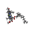
-Macromolecule #5: BACTERIOCHLOROPHYLL A
| Macromolecule | Name: BACTERIOCHLOROPHYLL A / type: ligand / ID: 5 / Number of copies: 48 / Formula: BCL |
|---|---|
| Molecular weight | Theoretical: 911.504 Da |
| Chemical component information |  ChemComp-BCL: |
-Macromolecule #6: LYCOPENE
| Macromolecule | Name: LYCOPENE / type: ligand / ID: 6 / Number of copies: 16 / Formula: LYC |
|---|---|
| Molecular weight | Theoretical: 536.873 Da |
| Chemical component information |  ChemComp-LYC: |
-Experimental details
-Structure determination
| Method | cryo EM |
|---|---|
 Processing Processing | single particle reconstruction |
| Aggregation state | particle |
- Sample preparation
| Buffer | pH: 7.5 |
|---|---|
| Vitrification | Cryogen name: ETHANE |
- Electron microscopy
| Microscope | FEI TALOS ARCTICA |
|---|---|
| Image recording | Film or detector model: FEI FALCON III (4k x 4k) / Average electron dose: 54.56 e/Å2 |
| Electron beam | Acceleration voltage: 200 kV / Electron source:  FIELD EMISSION GUN FIELD EMISSION GUN |
| Electron optics | Illumination mode: FLOOD BEAM / Imaging mode: BRIGHT FIELD / Nominal defocus max: 3.4 µm / Nominal defocus min: 1.0 µm |
| Experimental equipment |  Model: Talos Arctica / Image courtesy: FEI Company |
 Movie
Movie Controller


 Z (Sec.)
 Y (Row.)
 X (Col.)


