National Institutes of Health/National Institute of Environmental Health Sciences (NIH/NIEHS)
ZIA ES 103341
United States
National Institutes of Health/National Cancer Institute (NIH/NCI)
ZIA BC 011943
United States
National Institutes of Health/National Institute of Environmental Health Sciences (NIH/NIEHS)
ZIC ES 10326
United States
National Institutes of Health/National Cancer Institute (NIH/NCI)
ZIC BC 011891
United States
National Institutes of Health/National Cancer Institute (NIH/NCI)
Z01 BC010891
United States
National Institutes of Health/National Cancer Institute (NIH/NCI)
ZIA BC010891
United States
National Institutes of Health/National Cancer Institute (NIH/NCI)
75N91019D00024
United States
Citation
Journal: bioRxiv / Year: 2021 Title: Camel nanobodies broadly neutralize SARS-CoV-2 variants. Authors: Jessica Hong / Hyung Joon Kwon / Raul Cachau / Catherine Z Chen / Kevin John Butay / Zhijian Duan / Dan Li / Hua Ren / Tianyuzhou Liang / Jianghai Zhu / Venkata P Dandey / Negin Martin / ...Authors: Jessica Hong / Hyung Joon Kwon / Raul Cachau / Catherine Z Chen / Kevin John Butay / Zhijian Duan / Dan Li / Hua Ren / Tianyuzhou Liang / Jianghai Zhu / Venkata P Dandey / Negin Martin / Dominic Esposito / Uriel Ortega-Rodriguez / Miao Xu / Mario J Borgnia / Hang Xie / Mitchell Ho / Abstract: With the emergence of SARS-CoV-2 variants, there is urgent need to develop broadly neutralizing antibodies. Here, we isolate two V H nanobodies (7A3 and 8A2) from dromedary camels by phage display, ...With the emergence of SARS-CoV-2 variants, there is urgent need to develop broadly neutralizing antibodies. Here, we isolate two V H nanobodies (7A3 and 8A2) from dromedary camels by phage display, which have high affinity for the receptor-binding domain (RBD) and broad neutralization activities against SARS-CoV-2 and its emerging variants. Cryo-EM complex structures reveal that 8A2 binds the RBD in its up mode and 7A3 inhibits receptor binding by uniquely targeting a highly conserved and deeply buried site in the spike regardless of the RBD conformational state. 7A3 at a dose of ≥5 mg/kg efficiently protects K18-hACE2 transgenic mice from the lethal challenge of B.1.351 or B.1.617.2, suggesting that the nanobody has promising therapeutic potentials to curb the COVID-19 surge with emerging SARS-CoV-2 variants. ONE-SENTENCE SUMMARY: Dromedary camel ( ) V H phage libraries were built for isolation of the nanobodies that broadly neutralize SARS-CoV-2 variants.
Model: Homemade / Material: GOLD / Mesh: 300 / Support film - Material: GOLD / Support film - topology: HOLEY / Support film - Film thickness: 30 / Pretreatment - Type: PLASMA CLEANING / Pretreatment - Time: 75 sec. / Pretreatment - Atmosphere: OTHER
Vitrification
Cryogen name: ETHANE / Chamber humidity: 95 % / Chamber temperature: 298 K / Instrument: LEICA EM GP
Details
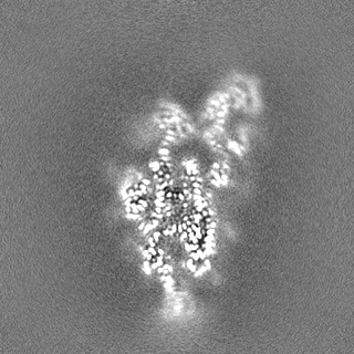
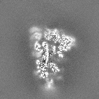
Complexes of spike with 7A3 and/or 8A2 VHHs were prepared by mixing the components at a spike trimer to nanobody molar ratio of 1:6. The final concentration of spike trimer was 3 uM in PBS at pH 7 with the addition of 5 mM imidazole. Because the 8A2 stock was too diluted, complexes involving this nanobody were prepared at 0.5 uM spike trimer followed by a 6-fold concentration using a 10 kDa cutoff centrifugal filter (Amicon Ultra). All complexes were incubated on ice for at least 5 min prior to grid preparation.
-
Electron microscopy #1
Microscopy ID
1
Microscope
FEI TALOS ARCTICA
Image recording
Image recording ID: 1 / Film or detector model: GATAN K2 SUMMIT (4k x 4k) / Detector mode: COUNTING / Average exposure time: 1.0 sec. / Average electron dose: 60.0 e/Å2
Electron beam
Acceleration voltage: 200 kV / Electron source: FIELD EMISSION GUN
In the structure databanks used in Yorodumi, some data are registered as the other names, "COVID-19 virus" and "2019-nCoV". Here are the details of the virus and the list of structure data.
Jan 31, 2019. EMDB accession codes are about to change! (news from PDBe EMDB page)
EMDB accession codes are about to change! (news from PDBe EMDB page)
The allocation of 4 digits for EMDB accession codes will soon come to an end. Whilst these codes will remain in use, new EMDB accession codes will include an additional digit and will expand incrementally as the available range of codes is exhausted. The current 4-digit format prefixed with “EMD-” (i.e. EMD-XXXX) will advance to a 5-digit format (i.e. EMD-XXXXX), and so on. It is currently estimated that the 4-digit codes will be depleted around Spring 2019, at which point the 5-digit format will come into force.
The EM Navigator/Yorodumi systems omit the EMD- prefix.
Related info.:Q: What is EMD? / ID/Accession-code notation in Yorodumi/EM Navigator
Yorodumi is a browser for structure data from EMDB, PDB, SASBDB, etc.
This page is also the successor to EM Navigator detail page, and also detail information page/front-end page for Omokage search.
The word "yorodu" (or yorozu) is an old Japanese word meaning "ten thousand". "mi" (miru) is to see.
Related info.:EMDB / PDB / SASBDB / Comparison of 3 databanks / Yorodumi Search / Aug 31, 2016. New EM Navigator & Yorodumi / Yorodumi Papers / Jmol/JSmol / Function and homology information / Changes in new EM Navigator and Yorodumi
 Movie
Movie Controller

 Yorodumi
 Open data
 Basic information
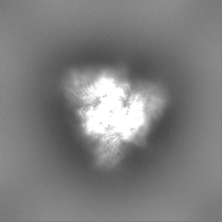
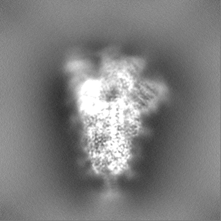
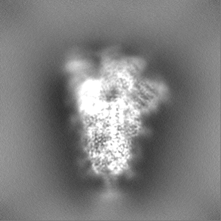
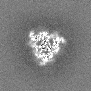
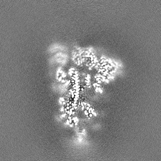
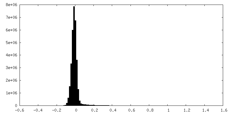
 Map data
 Sample
 Keywords
 Function and homology information
 Severe acute respiratory syndrome coronavirus 2 /
 Authors
 United States, 7 items
  Citation
 Structure visualization
 Downloads & links
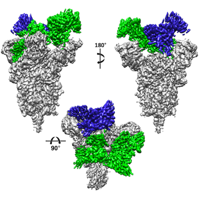 emd_26062.png
 http://ftp.pdbj.org/pub/emdb/structures/EMD-26062
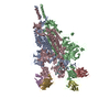


 Z (Sec.)
 Y (Row.)
 X (Col.)


 Sample components
 Homo sapiens (human)
 Processing
 Electron microscopy #1
 FIELD EMISSION GUN

