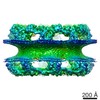
- EMDB-25817: Cryo-EM map of protomer of the cytoplasmic ring of the nuclear po... -
+
Open data
ID or keywords:
Loading...
-
Basic information
Entry
Database: EMDB / ID: EMD-25817
Title
Cryo-EM map of protomer of the cytoplasmic ring of the nuclear pore complex from Xenopus laevis
Map data
Cryo-EM map of protomer of the cytoplasmic ring of the Nuclear pore complex from Xenopus laevis.
Sample
Complex: Cytoplasmic ring of Nuclear Pore Complex
Keywords
Nuclear pore complex / NUCLEAR PROTEIN
Function / homology
Function and homology information
: / nitrogen compound transport / macromolecule localization / system development / nephron development / GATOR2 complex / macromolecule metabolic process / Seh1-associated complex / nuclear pore inner ring / animal organ development ...: / nitrogen compound transport / macromolecule localization / system development / nephron development / GATOR2 complex / macromolecule metabolic process / Seh1-associated complex / nuclear pore inner ring / animal organ development / COPII-coated vesicle budding / protein exit from endoplasmic reticulum / nuclear pore central transport channel / transcription-dependent tethering of RNA polymerase II gene DNA at nuclear periphery / nuclear pore outer ring / protein localization to nuclear inner membrane / nuclear pore organization / COPII vesicle coat / post-transcriptional tethering of RNA polymerase II gene DNA at nuclear periphery / attachment of mitotic spindle microtubules to kinetochore / structural constituent of nuclear pore / RNA export from nucleus / nucleocytoplasmic transport / nuclear localization sequence binding / poly(A)+ mRNA export from nucleus / mitotic metaphase chromosome alignment / nuclear pore / ribosomal large subunit export from nucleus / positive regulation of TOR signaling / mRNA transport / mRNA export from nucleus / ribosomal small subunit export from nucleus / positive regulation of TORC1 signaling / cellular response to nutrient levels / cellular response to amino acid starvation / GTPase activator activity / chromosome segregation / phospholipid binding / protein import into nucleus / kinetochore / spindle pole / protein transport / nuclear membrane / cell division / lysosomal membrane / centrosome / positive regulation of DNA-templated transcription / structural molecule activity / DNA binding / zinc ion binding / cytosol / cytoplasm Similarity search - Function
Journal: Science / Year: 2022 Title: Structure of cytoplasmic ring of nuclear pore complex by integrative cryo-EM and AlphaFold. Authors: Pietro Fontana / Ying Dong / Xiong Pi / Alexander B Tong / Corey W Hecksel / Longfei Wang / Tian-Min Fu / Carlos Bustamante / Hao Wu / Abstract: INTRODUCTION The nuclear pore complex (NPC) is the molecular conduit in the nuclear membrane of eukaryotic cells that regulates import and export of biomolecules between the nucleus and the cytosol, ...INTRODUCTION The nuclear pore complex (NPC) is the molecular conduit in the nuclear membrane of eukaryotic cells that regulates import and export of biomolecules between the nucleus and the cytosol, with vertebrate NPCs ~110 to 125 MDa in molecular mass and ~120 nm in diameter. NPCs are organized into four main rings: the cytoplasmic ring (CR) at the cytosolic side, the inner ring and the luminal ring on the plane of the nuclear membrane, and the nuclear ring facing the nucleus. Each ring possesses an approximate eightfold symmetry and is composed of multiple copies of different nucleoporins. NPCs have been implicated in numerous biological processes, and their dysfunctions are associated with a growing number of serious human diseases. However, despite pioneering studies from many groups over the past two decades, we still lack a full understanding of NPCs' organization, dynamics, and complexity. RATIONALE We used the oocyte as a model system for the structural characterization because each oocyte possesses a large number of NPC particles that can be visualized on native nuclear membranes without the aid of detergent extraction. We used single-particle cryo-electron microscopy (cryo-EM) analysis on data collected at different stage tilt angles for three-dimensional reconstruction and structure prediction with AlphaFold for model building. RESULTS We reconstructed the CR map of NPC at 6.9 and 6.7 Å resolutions for the full CR protomer and a core region, respectively, and predicted the structures of the individual nucleoporins using AlphaFold because no high-resolution models of Nups were available. For any ambiguous subunit interactions, we also predicted complex structures, which further guided model fitting of the CR protomer. We placed the nucleoporin or complex structures into the CR density to obtain an almost full CR atomic model, composed of the inner and outer Y-complexes, two copies of Nup205, two copies of the Nup214-Nup88-Nup62 complex, one Nup155, and five copies of Nup358. In particular, we predicted the largest protein in the NPC, Nup358, as having an S-shaped globular domain, a coiled-coil domain, and a largely disordered C-terminal region containing phenylalanine-glycine (FG) repeats previously shown to form a gel-like condensate phase for selective cargo passage. Four of the Nup358 copies clamp around the inner and outer Y-complexes to stabilize the CR, and the fifth Nup358 situates in the center of the cluster of clamps. AlphaFold also predicted a homo-oligomeric, likely specifically pentameric, coiled-coil structure of Nup358 that may provide the avidity for Nup358 recruitment to the NPC and for lowering the threshold for Nup358 condensation in NPC biogenesis. CONCLUSION Our studies offer an example of integrative cryo-EM and structure prediction as a general approach for attaining more precise models of megadalton protein complexes from medium-resolution density maps. The more accurate and almost complete model of the CR presented here expands our understanding of the molecular interactions in the NPC and represents a substantial step forward toward the molecular architecture of a full NPC, with implications for NPC function, biogenesis, and regulation. [Figure: see text].
In the structure databanks used in Yorodumi, some data are registered as the other names, "COVID-19 virus" and "2019-nCoV". Here are the details of the virus and the list of structure data.
Jan 31, 2019. EMDB accession codes are about to change! (news from PDBe EMDB page)
EMDB accession codes are about to change! (news from PDBe EMDB page)
The allocation of 4 digits for EMDB accession codes will soon come to an end. Whilst these codes will remain in use, new EMDB accession codes will include an additional digit and will expand incrementally as the available range of codes is exhausted. The current 4-digit format prefixed with “EMD-” (i.e. EMD-XXXX) will advance to a 5-digit format (i.e. EMD-XXXXX), and so on. It is currently estimated that the 4-digit codes will be depleted around Spring 2019, at which point the 5-digit format will come into force.
The EM Navigator/Yorodumi systems omit the EMD- prefix.
Related info.:Q: What is EMD? / ID/Accession-code notation in Yorodumi/EM Navigator
Yorodumi is a browser for structure data from EMDB, PDB, SASBDB, etc.
This page is also the successor to EM Navigator detail page, and also detail information page/front-end page for Omokage search.
The word "yorodu" (or yorozu) is an old Japanese word meaning "ten thousand". "mi" (miru) is to see.
Related info.:EMDB / PDB / SASBDB / Comparison of 3 databanks / Yorodumi Search / Aug 31, 2016. New EM Navigator & Yorodumi / Yorodumi Papers / Jmol/JSmol / Function and homology information / Changes in new EM Navigator and Yorodumi
 Movie
Movie Controller

 Yorodumi
 Open data
 Basic information
 Map data
 Sample
 Keywords
 Function and homology information
 Authors
 Citation
 Structure visualization
 Downloads & links
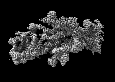 emd_25817.png
 http://ftp.pdbj.org/pub/emdb/structures/EMD-25817


 Z (Sec.)
 Y (Row.)
 X (Col.)


 Sample components
 Processing
 Electron microscopy
 FIELD EMISSION GUN