 Movie
Movie Controller


+ Open data

- Basic information
| Entry |  | |||||||||
|---|---|---|---|---|---|---|---|---|---|---|
| Title | Structure of bacteriophage lambda tube protein V in C3 | |||||||||
 Map data Map data | Bacteriophage lambda tube protein V in C3 | |||||||||
 Sample Sample |
| |||||||||
 Keywords Keywords | Virus / Phage / Lambda / Siphoviridae / Tail / Tube | |||||||||
| Function / homology |  Function and homology information Function and homology informationvirus tail, tube / symbiont genome ejection through host cell envelope, long flexible tail mechanism / viral tail assembly / host cell cytoplasm Similarity search - Function | |||||||||
| Biological species |  Escherichia virus Lambda Escherichia virus Lambda | |||||||||
| Method | single particle reconstruction / cryo EM / Resolution: 2.7 Å | |||||||||
 Authors Authors | Prokhorov NS / Yang Q | |||||||||
| Funding support |  United States, 1 items United States, 1 items
| |||||||||
 Citation Citation | Journal: To Be Published Title: Structure of bacteriophage lambda tube protein V in C6 Authors: Prokhorov NS / Yang Q / Catalano CE / Morais MC | |||||||||
| History |
|
- Structure visualization
| Supplemental images |
|---|

- Downloads & links
-EMDB archive
| Map data | emd_25675.map.gz | 54.1 MB | EMDB map data format | |
|---|---|---|---|---|
| Header (meta data) | emd-25675-v30.xmlemd-25675.xml | 13.2 KB 13.2 KB | Display Display | EMDB header |
| FSC (resolution estimation) | emd_25675_fsc.xml | 11.6 KB | Display | FSC data file |
| Images | 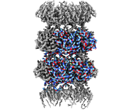 emd_25675.png emd_25675.png | 124.5 KB | ||
| Filedesc metadata | emd-25675.cif.gz | 5 KB | ||
| Others | emd_25675_half_map_1.map.gzemd_25675_half_map_2.map.gz | 58.8 MB 58.8 MB | ||
| Archive directory |  http://ftp.pdbj.org/pub/emdb/structures/EMD-25675ftp://ftp.pdbj.org/pub/emdb/structures/EMD-25675 http://ftp.pdbj.org/pub/emdb/structures/EMD-25675ftp://ftp.pdbj.org/pub/emdb/structures/EMD-25675 | HTTPS FTP |
-Related structure data
| Related structure data | 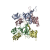 7t4fMC 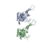 7t2eC C: citing same article ( M: atomic model generated by this map |
|---|---|
| Similar structure data |

-Links
| EMDB pages | EMDB (EBI/PDBe) / EMDataResource |
|---|
-Map
| File | Download / File: emd_25675.map.gz / Format: CCP4 / Size: 64 MB / Type: IMAGE STORED AS FLOATING POINT NUMBER (4 BYTES) | ||||||||||||||||||||||||||||||||||||
|---|---|---|---|---|---|---|---|---|---|---|---|---|---|---|---|---|---|---|---|---|---|---|---|---|---|---|---|---|---|---|---|---|---|---|---|---|---|
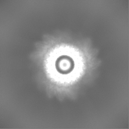
| Annotation | Bacteriophage lambda tube protein V in C3 | ||||||||||||||||||||||||||||||||||||
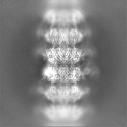
| Projections & slices | Image control
Images are generated by Spider. | ||||||||||||||||||||||||||||||||||||
| Voxel size | X=Y=Z: 1.1 Å | ||||||||||||||||||||||||||||||||||||
| Density |
| ||||||||||||||||||||||||||||||||||||
| Symmetry | Space group: 1 | ||||||||||||||||||||||||||||||||||||
| Details | EMDB XML:
|
 X (Sec.)
 Y (Row.)
 Z (Col.)


-Supplemental data
-Half map: Bacteriophage lambda tube protein V in C3
| File | emd_25675_half_map_1.map | ||||||||||||
|---|---|---|---|---|---|---|---|---|---|---|---|---|---|
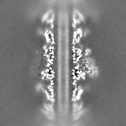
| Annotation | Bacteriophage lambda tube protein V in C3 | ||||||||||||
| Projections & Slices |
| ||||||||||||
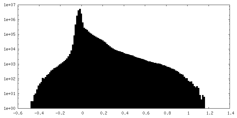
| Density Histograms |


-Half map: Bacteriophage lambda tube protein V in C3
| File | emd_25675_half_map_2.map | ||||||||||||
|---|---|---|---|---|---|---|---|---|---|---|---|---|---|
| Annotation | Bacteriophage lambda tube protein V in C3 | ||||||||||||
| Projections & Slices |
| ||||||||||||
| Density Histograms |


- Sample components
-Entire : Escherichia virus Lambda
| Entire | Name: Escherichia virus Lambda |
|---|---|
| Components |
|
-Supramolecule #1: Escherichia virus Lambda
| Supramolecule | Name: Escherichia virus Lambda / type: virus / ID: 1 / Parent: 0 / Macromolecule list: all / NCBI-ID: 10710 / Sci species name: Escherichia virus Lambda / Virus type: VIRION / Virus isolate: STRAIN / Virus enveloped: No / Virus empty: No |
|---|
-Macromolecule #1: Tail tube protein
| Macromolecule | Name: Tail tube protein / type: protein_or_peptide / ID: 1 / Number of copies: 4 / Enantiomer: LEVO |
|---|---|
| Source (natural) | Organism: Escherichia virus Lambda |
| Molecular weight | Theoretical: 25.700582 KDa |
| Recombinant expression | Organism:  |
| Sequence | String: PVPNPTMPVK GAGTTLWVYK GSGDPYANPL SDVDWSRLAK VKDLTPGELT AESYDDSYLD DEDADWTATG QGQKSAGDTS FTLAWMPGE QGQQALLAWF NEGDTRAYKI RFPNGTVDVF RGWVSSIGKA VTAKEVITRT VKVTNVGRPS MAEDRSTVTA A TGMTVTPA ...String: PVPNPTMPVK GAGTTLWVYK GSGDPYANPL SDVDWSRLAK VKDLTPGELT AESYDDSYLD DEDADWTATG QGQKSAGDTS FTLAWMPGE QGQQALLAWF NEGDTRAYKI RFPNGTVDVF RGWVSSIGKA VTAKEVITRT VKVTNVGRPS MAEDRSTVTA A TGMTVTPA STSVVKGQST TLTVAFQPEG VTDKSFRAVS ADKTKATVSV SGMTITVNGV AAGKVNIPVV SGNGEFAAVA EI TVTAS UniProtKB: Tail tube protein |
-Experimental details
-Structure determination
| Method | cryo EM |
|---|---|
 Processing Processing | single particle reconstruction |
| Aggregation state | particle |
-Sample preparation
| Buffer | pH: 7.5 |
|---|---|
| Vitrification | Cryogen name: ETHANE |
- Electron microscopy
| Microscope | FEI TITAN KRIOS |
|---|---|
| Image recording | Film or detector model: GATAN K3 BIOQUANTUM (6k x 4k) / Average electron dose: 35.6 e/Å2 |
| Electron beam | Acceleration voltage: 300 kV / Electron source:  FIELD EMISSION GUN FIELD EMISSION GUN |
| Electron optics | Illumination mode: FLOOD BEAM / Imaging mode: BRIGHT FIELD / Nominal defocus max: 3.0 µm / Nominal defocus min: 1.0 µm |
| Experimental equipment |  Model: Titan Krios / Image courtesy: FEI Company |

